Mohs micrographic surgery (first stage up to 5 lesions) Dermatology Surgery
Mohs micrographic surgery is a precise method to remove certain skin cancers.
Overview
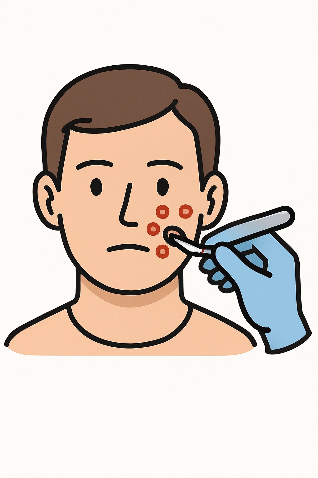
Mohs micrographic surgery is a precise method to remove certain skin cancers. The surgeon removes a thin layer of tissue, maps it, and examines it under a microscope on the same day. If cancer cells are still seen at the edge, another thin layer is taken only from the area where cancer remains. The "first stage" is the initial round of removal and analysis. Many people need more than one stage at the same visit. This approach aims to fully remove the cancer while preserving as much healthy skin as possible, which is helpful on areas like the face, ears, hands, feet, and genitals.
Also known as: Mohs surgery, Micrographic surgery, Mohs
Preparation & Next Steps
Everything you need to know before and after your procedure
Before Care
- Plan to be at the clinic for several hours in case multiple stages are needed the same day
- Provide a current list of medicines and supplements, including blood thinners, to the clinic
- Confirm eating or fasting instructions with the clinic; most Mohs procedures use local anesthesia
- Wear comfortable clothing and avoid makeup, lotions, or sunscreen on the area to be treated if accessible
- Arrange a ride if a large repair is possible or if you prefer not to drive after the visit
- Set work and caregiving plans in case stitches or a bandage limit activity
- Bring a list of allergies and any prior reactions to anesthesia or antiseptics
- Follow any clinic guidance about washing the area or shaving before arrival
- Bring a book or phone for waiting periods between microscope checks
After Care
- Keep the pressure bandage and wound clean and dry as instructed by your clinic
- Use pain control methods recommended by your clinical team and avoid picking at the site
- Limit bending, heavy lifting, and strenuous exercise that could cause bleeding until the clinic says activity is safe
- Elevate the area and use cold packs wrapped in a cloth to help reduce swelling if advised by the clinic
- Watch for increasing redness, warmth, swelling, drainage, or fever and contact the clinic if these appear
- Follow instructions for dressing changes, ointments, and when showering is allowed
- Protect the area from sun with clothing or shade while healing
- Attend scheduled follow-up for wound check and stitch removal if used
- If a flap or graft was placed, monitor the site as instructed and report concerns to the clinic
- Keep the clinic’s after-hours contact information handy for urgent wound questions
Clinical Information
Important medical details about this procedure
Indications
- Basal cell carcinoma
- Squamous cell carcinoma
- Skin cancers on the face, ears, eyelids, nose, lips, hands, feet, or genitals where tissue sparing matters
- Recurrent tumors after prior treatment
- Tumors with ill-defined borders or aggressive growth patterns
- Selected cases of melanoma in situ using specialized techniques
Alternatives
- Standard surgical excision
- Curettage and electrodesiccation (scraping and cautery)
- Cryosurgery (freezing)
- Radiation therapy
- Topical medicines for certain superficial tumors (for example, imiquimod or 5-fluorouracil)
- Photodynamic therapy for some superficial lesions
Risks
- Bleeding or bruising
- Infection
- Pain, swelling, or discomfort at the site
- Scar or changes in skin color or texture
- Numbness or nerve injury affecting sensation or movement
- Damage to nearby structures (for example, eyelid, lip, or nose)
- Need for stitches, flap, or skin graft to close the wound
- Allergic reaction to local anesthesia or antiseptics
- Cancer recurrence that may need more treatment
Contraindications
- Severe medical instability that makes an office-based procedure unsafe
- Inability to cooperate or remain still for a local anesthetic procedure
- History of severe allergy to local anesthetics without acceptable alternatives
- Uncontrolled bleeding disorder
- Active skin infection at the site that requires initial treatment
Recovery Timeline
What to expect during your recovery
Most people resume light daily activities soon after surgery. Stitches, if used, are often removed within about 5 to 14 days depending on location. The scar continues to mature over several months.
Typical Range
7–14 days
Return to Work
1–3 days
Recovery Milestones
Go home the same day; do light indoor activities
Return to desk work if comfortable and if job demands are light
Increase light walking and daily tasks; avoid heavy lifting and strenuous exercise
Stitch removal in clinic if stitches were placed
Gradual return to full activity as the wound continues to strengthen
Scar appearance continues to mature over months
Frequently Asked Questions
Common questions and expert answers about this procedure
What is Mohs micrographic surgery?
What is Mohs micrographic surgery?
It is a stepwise skin cancer treatment where thin layers are removed and checked under a microscope the same day until no cancer remains at the edges.
What does the first stage mean?
What does the first stage mean?
The first stage is the initial layer of tissue removal and analysis. If cancer cells are still at the edge, more stages are done the same day.
How long does Mohs surgery take?
How long does Mohs surgery take?
It varies. Each stage includes removal, processing, and microscopic review, which can take an hour or more. Some people need several stages.
Will I be awake during the procedure?
Will I be awake during the procedure?
Yes. Mohs surgery is usually done with local anesthesia. You remain awake, and the area is numbed.
Why choose Mohs surgery?
Why choose Mohs surgery?
It checks 100% of the surgical margin and removes as little normal skin as possible, which is helpful for cancers in sensitive or high-risk areas.
Will I have a scar?
Will I have a scar?
Any skin surgery can leave a scar. The goal is to remove the cancer and close the wound in a way that supports healing and appearance.
How is the wound closed?
How is the wound closed?
Depending on size and location, it may be closed with stitches, a skin flap, a skin graft, or left to heal on its own.
Who performs Mohs surgery?
Who performs Mohs surgery?
A dermatologist with specialized training in Mohs micrographic surgery typically performs it and reviews the tissues under the microscope.
References
Medical literature and sources