Ankle X-ray (complete ≥3 views) Diagnostic Imaging
An ankle X-ray is a quick imaging test that uses a small amount of radiation to make pictures of the bones in your ankle.
Overview
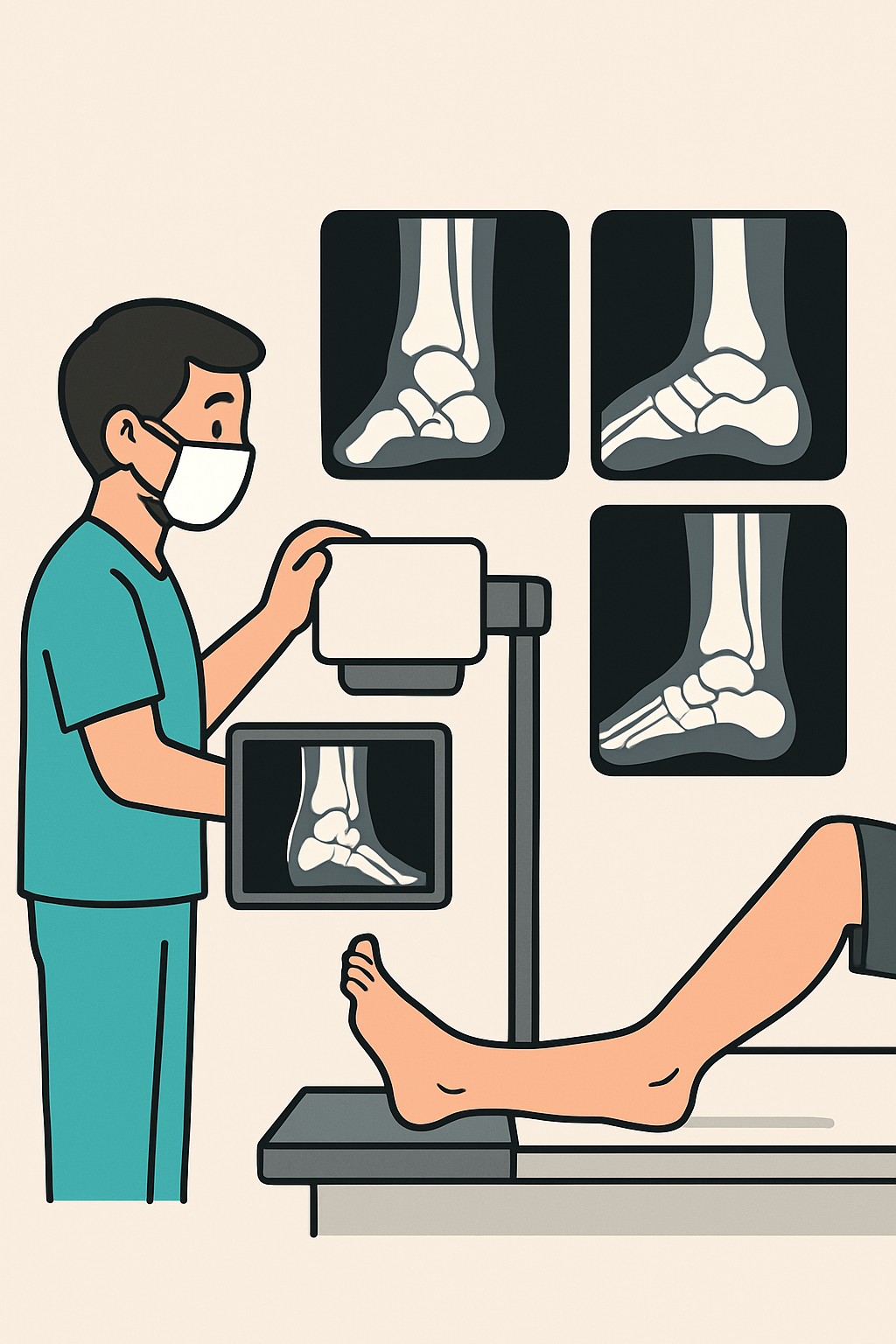
An ankle X-ray is a quick imaging test that uses a small amount of radiation to make pictures of the bones in your ankle. A complete series uses three or more views taken from different angles to help find problems that might not show on a single view. Clinicians order this test to look for broken bones, dislocations, joint alignment issues, and signs of arthritis or infection. Multiple views improve the chance of seeing small or complex injuries.
Also known as: Ankle radiograph, Ankle series, Ankle X-ray 3 views, Ankle plain films
Preparation & Next Steps
Everything you need to know before and after your procedure
Before Care
- Bring your photo ID, insurance card, and the imaging order if you have it
- Wear clothing that allows easy access to the ankle or expect to change into a gown
- Remove jewelry or metal near the ankle that could obscure the images
- Tell the imaging staff if you might be pregnant or are breastfeeding
- Share recent ankle injuries, surgeries, or previous imaging with the technologist
- Bring or upload prior ankle images for comparison if available
- Arrive a little early for check-in and safety screening
- Use a brace, boot, cane, or crutches as needed for safe mobility
- No fasting or sedation is typically required
After Care
- Resume normal activities unless your clinician has given you other instructions
- Use your usual mobility aids to avoid strain on the ankle
- Check your patient portal or plan for how you will receive the report
- Keep copies of your images and report for future reference
- Ask the ordering clinic about timing for results and any next steps
- Know that some injuries do not appear right away; follow-up imaging may be ordered
- Contact your clinician if pain, swelling, or function worsens after the exam
- Track symptoms so changes can be compared with imaging results
Clinical Information
Important medical details about this procedure
Indications
- Pain, swelling, or tenderness after a fall or twist
- Suspected ankle fracture or dislocation
- Inability to bear weight after an injury
- Evaluation of chronic ankle pain or arthritis
- Follow-up on a known fracture or post-surgery hardware
- Suspected bone infection or bone tumor
- Checking alignment after reduction or casting
Alternatives
- Observation and physical exam when appropriate
- Ultrasound for soft tissue concerns
- CT scan for complex fractures or pre-surgical planning
- MRI for ligament, tendon, cartilage, or occult injuries
- Bone scan for certain infections or stress injuries
Risks
- Low radiation exposure from the X-ray
- Rare need for repeat imaging if views are unclear
- Discomfort from positioning the ankle
- Missed subtle injuries that may need follow-up imaging
Contraindications
- Known or possible pregnancy without prior discussion of shielding or alternatives
- Inability to remain still for image capture without safe support
Recovery Timeline
What to expect during your recovery
There is no medical recovery period from a standard ankle X-ray. Most people continue their usual activities immediately after the exam.
Typical Range
Same day
Return to Work
Same day
Recovery Milestones
Resume normal daily activities
Access images or report via portal if available
Complete any follow-up imaging if ordered
Frequently Asked Questions
Common questions and expert answers about this procedure
What does complete ≥3 views mean?
What does complete ≥3 views mean?
It means the technologist takes at least three pictures from different angles. This helps reveal fractures or alignment issues that a single view might miss.
How long does an ankle X-ray take?
How long does an ankle X-ray take?
The imaging usually takes about 10 to 15 minutes. Extra time may be needed for check-in and positioning.
Will the X-ray hurt?
Will the X-ray hurt?
The X-ray itself is painless. Positioning the ankle may be uncomfortable if you are injured, but it is brief.
Is there radiation exposure?
Is there radiation exposure?
Yes, but it is a low dose for a standard ankle X-ray. The benefits of diagnosing a fracture or other problem usually outweigh the small risk.
Is it safe during pregnancy?
Is it safe during pregnancy?
Many diagnostic X-rays expose the fetus to very low doses. Shielding and alternative imaging may be considered depending on the situation.
Do I need to prepare or fast?
Do I need to prepare or fast?
No special preparation or fasting is typical. You may be asked to remove metal objects near the ankle.
When will I get results?
When will I get results?
A radiologist interprets the images and sends a report to the ordering clinician. Timing varies by facility and urgency.
What if my X-ray is normal but I still have pain?
What if my X-ray is normal but I still have pain?
Some injuries involve soft tissues or are hard to see early on. Your clinician may use exam findings and, if needed, other imaging.
References
Medical literature and sources