Chest X-ray (2 views - portable film) Diagnostic Imaging
A chest X-ray uses a small amount of radiation to create images of the heart, lungs, airways, and bones of the chest.
Overview
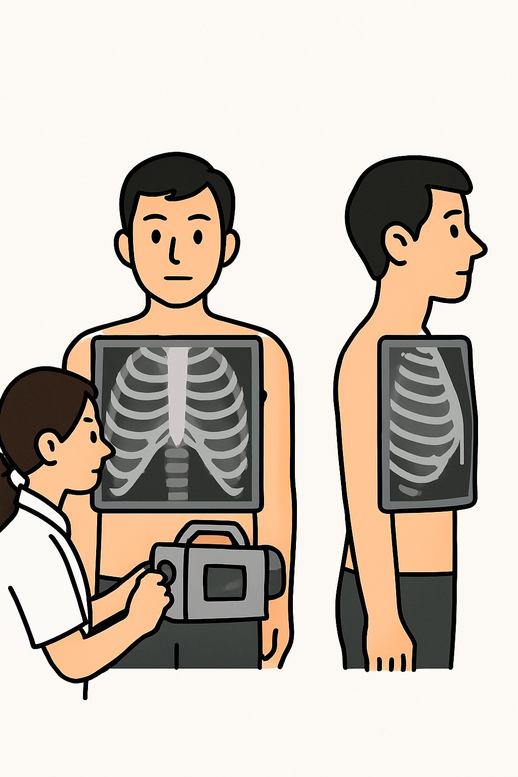
A chest X-ray uses a small amount of radiation to create images of the heart, lungs, airways, and bones of the chest. A portable exam means the X-ray machine comes to the patient, often at the bedside in a hospital or care facility. Two views are taken from different angles to give a clearer picture and help find issues that may not show on a single view. Clinicians use this test to look for pneumonia, fluid, a collapsed lung, heart enlargement, broken ribs, and to check the position of devices like tubes or lines.
Also known as: Portable chest radiograph (2 views), Portable CXR (2 views), Bedside chest X-ray
Preparation & Next Steps
Everything you need to know before and after your procedure
Before Care
- Bring the imaging order and a photo ID if you are coming from outside a hospital unit
- Tell the technologist if you might be pregnant so shielding can be used when appropriate
- Remove necklaces, body jewelry, or other metal near the chest and neck if possible
- Wear a loose, easy-to-remove top; you may be asked to change into a gown
- Be ready to sit upright or lie flat; two different positions or angles are usually needed
- Let staff know about pain or limited movement so they can help position you comfortably
- Have recent chest imaging dates and locations available to help with comparison
- Keep oxygen, IV lines, or monitors in place; staff will position equipment safely
- Follow any facility infection-control instructions for the room and equipment
- Ask how and when results will be shared (patient portal, bedside team, or clinic call)
After Care
- Resume normal activities and eating right away unless your care team advises otherwise for other reasons
- Stay available briefly in case an extra view is requested for clarity
- If the X-ray was to check a tube or line, staff may adjust the device based on the image
- Review your report and images in the patient portal when released
- Keep a personal record of your imaging history and where studies were performed
- Contact your clinician if your breathing, chest pain, or other symptoms worsen
- Ask about next steps if the report mentions follow-up imaging or comparison with prior studies
- Use clean hands when handling any dressings or devices that were repositioned after imaging
Clinical Information
Important medical details about this procedure
Indications
- Cough, fever, or suspected pneumonia
- Shortness of breath or low oxygen levels
- Chest pain or trauma
- Monitoring heart size or fluid in the lungs (heart failure)
- Checking placement of feeding tubes, central lines, or endotracheal tubes
- Follow-up after chest procedures or surgery
- Evaluation of abnormal exam or lab findings
Alternatives
- Standard (non-portable) chest X-ray in an imaging room
- Chest CT for more detailed imaging when needed
- Lung ultrasound for certain conditions (such as fluid or pneumothorax)
- Observation and repeat exam when immediate imaging is not necessary
Risks
- Small exposure to ionizing radiation
- Image quality limits from motion or positioning may require repeat images
- Incidental findings that may lead to additional testing
- Temporary discomfort from holding certain positions
Contraindications
- No absolute contraindications for a standard chest X-ray
- Pregnancy is not a strict contraindication; shielding and justification are used
- Inability to remain still or follow simple breathing instructions may limit image quality
- Large dressings, monitoring equipment, or metal objects over the chest can obscure the image
Recovery Timeline
What to expect during your recovery
There is no medical recovery period for a chest X-ray. Most people return to normal activities immediately after the images are taken.
Typical Range
Same day
Return to Work
Same day
Recovery Milestones
Resume normal daily activities
Access the radiology report in the patient portal if available
Discuss results with your clinician if a follow-up plan is needed
Frequently Asked Questions
Common questions and expert answers about this procedure
What does portable chest X-ray mean?
What does portable chest X-ray mean?
The technologist brings a mobile X-ray unit to your bedside, often in a hospital or care facility, so you do not need to travel to the imaging department.
Why are two views taken?
Why are two views taken?
Images from different angles help show structures that can overlap on a single view and make it easier to spot problems.
How long does the test take?
How long does the test take?
Positioning and taking two images usually takes only a few minutes. Extra time may be needed in busy units or if additional views are requested.
Will I be exposed to a lot of radiation?
Will I be exposed to a lot of radiation?
A chest X-ray uses a small amount of radiation and is much lower than a CT scan. The benefit of finding important problems often outweighs this small risk.
Is it safe during pregnancy?
Is it safe during pregnancy?
Chest X-rays can be done in pregnancy when needed. Shielding and careful technique are used to limit exposure.
Do I need to fast or stop my medicines?
Do I need to fast or stop my medicines?
No fasting is needed, and medicines are usually continued. Follow any instructions from your care team for your overall treatment.
What can a chest X-ray show?
What can a chest X-ray show?
It can show pneumonia, fluid, a collapsed lung, enlarged heart, broken ribs, and device placement. Some issues need more detailed imaging like a CT.
How will I get my results?
How will I get my results?
A radiologist reads the images and sends a report to your clinician. Many facilities also post the report and images to the patient portal.
References
Medical literature and sources