Eye Exam with Retina Scan (Extended Ophthalmoscopy) Diagnostic Imaging
An eye exam with retina scan, also called extended ophthalmoscopy, is a detailed look at the back of the eye.
Overview
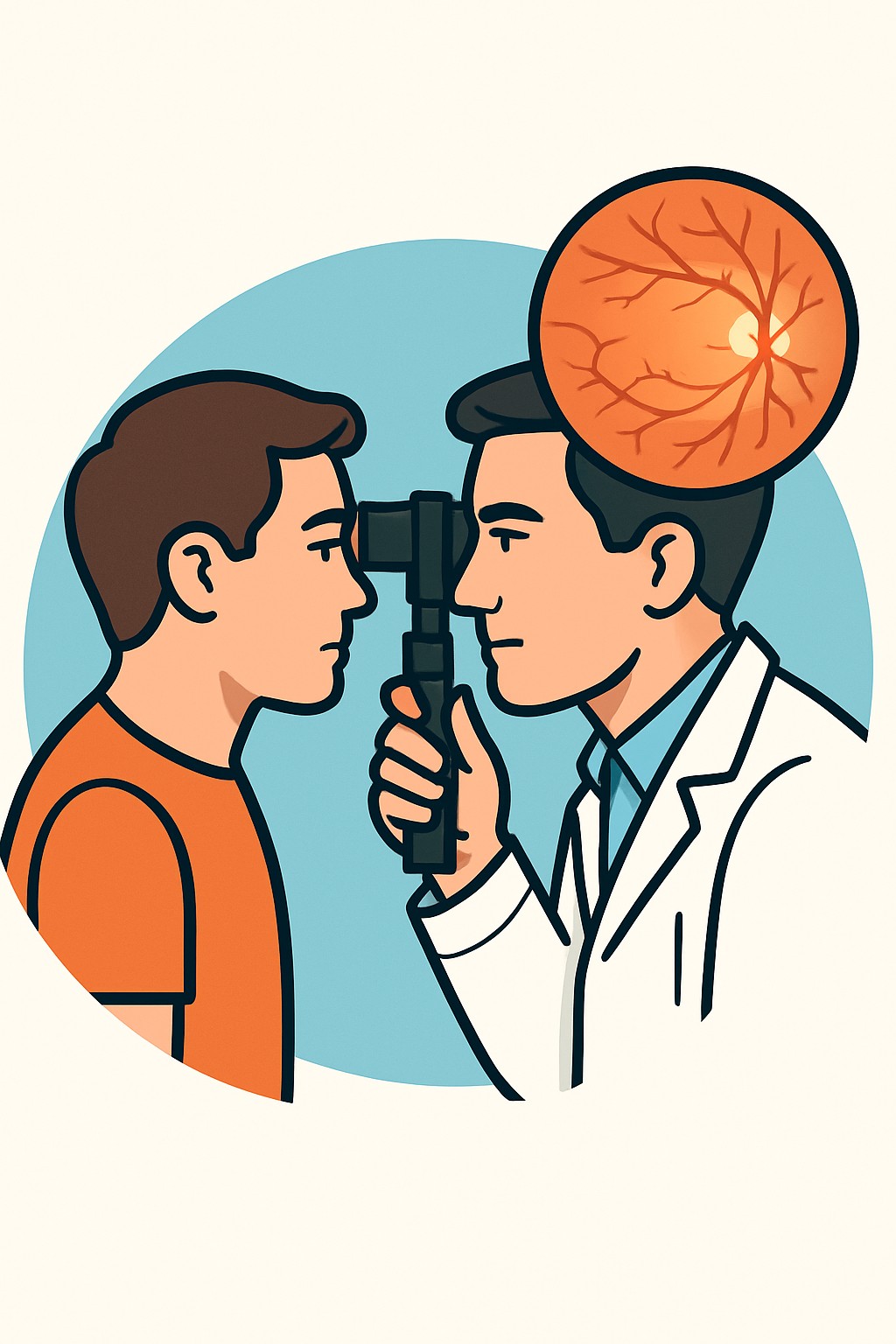
An eye exam with retina scan, also called extended ophthalmoscopy, is a detailed look at the back of the eye. The clinician usually puts in dilating drops to widen the pupils, then uses bright light and special lenses to view the retina and optic nerve. The retina is the light-sensing layer that lines the back of the eye, and the optic nerve carries visual signals to the brain. Retina scans may include digital retinal photos or optical coherence tomography (OCT), a non-contact imaging test that shows cross-sections of the retina. These tools help find or monitor eye diseases and explain symptoms like new floaters, flashes, or blurred vision.
Also known as: Dilated retinal exam, Dilated fundus exam, Retinal imaging, Comprehensive retinal exam
Preparation & Next Steps
Everything you need to know before and after your procedure
Before Care
- Bring your glasses, contact lens case, and a list of current medicines and eye drops
- Plan extra time after the visit because dilation can blur near vision for several hours
- Arrange transportation or be prepared to wait until your vision feels clear enough to drive
- Bring sunglasses to reduce light sensitivity after dilation
- Tell the staff about eye allergies, prior reactions to dilating drops, or glaucoma history
- Share any recent eye images or records, especially if you are seeing a new clinician
- Inform the clinic if you are pregnant or breastfeeding, particularly if dye-based imaging is being considered
- Avoid heavy scheduling right after the appointment in case your vision remains blurry
- Follow any clinic instructions about contact lens wear for imaging
- Have your pharmacy information ready in case eye drops are prescribed
After Care
- Expect light sensitivity and blurred near vision for several hours while the drops wear off
- Use sunglasses and reduce screen time until your vision feels comfortable
- Plan transportation if you do not feel safe driving while your pupils are dilated
- Follow the clinic’s instructions for any eye drops given during the visit
- Review your results and images in the patient portal or summary provided
- Schedule recommended follow-up or additional testing if advised by the clinic
- Resume contact lenses when your vision is clear and your eyes feel normal
- Contact a clinician if you develop severe eye pain, sudden vision loss, many new floaters or flashes, or a dark curtain in your vision
Clinical Information
Important medical details about this procedure
Indications
- Monitoring diabetes for diabetic retinopathy
- Evaluation of age-related macular degeneration
- New floaters, flashes of light, or a dark curtain in vision
- Blurry or distorted central vision
- High myopia with concern for retinal tears
- Eye trauma evaluation
- High blood pressure affecting the retina
- Inflammation inside the eye (uveitis)
- Follow-up after retinal surgery or treatment
Alternatives
- Standard dilated fundus exam without imaging
- Indirect ophthalmoscopy with headlamp and handheld lenses
- Digital fundus photography
- Optical coherence tomography (OCT)
- Ultra-widefield retinal imaging
- Teleophthalmology retinal photos for screening
- B-scan ocular ultrasound when the view is blocked by cataract or bleeding
Risks
- Temporary light sensitivity and blurred near vision from dilating drops
- Mild stinging with eye drops
- Rare allergic reaction to eye drops
- Short-term rise in eye pressure in people at risk for angle-closure glaucoma
- Temporary discomfort from bright lights or scleral depression during the exam
Contraindications
- Known hypersensitivity to dilating drops
- History or suspicion of narrow-angle glaucoma without prior assessment for dilation
- Recent eye surgery or trauma where dilation or pressure on the eye may be limited
- Active eye infection where contact procedures may be deferred
- Pregnancy considerations if dye-based imaging is planned
Recovery Timeline
What to expect during your recovery
There is no medical recovery period, but dilating drops can cause blurry near vision and light sensitivity for several hours. Most people resume normal activities the same day.
Typical Range
0–1 days
Return to Work
0–1 days
Recovery Milestones
Light sensitivity decreases as dilation wears off
Read and use screens as comfortable
Resume normal daily activities when vision feels clear
Frequently Asked Questions
Common questions and expert answers about this procedure
What is extended ophthalmoscopy?
What is extended ophthalmoscopy?
It is a detailed, dilated exam of the retina and optic nerve using bright light and special lenses. It may be documented with drawings or images.
Is a retina scan the same as a dilated exam?
Is a retina scan the same as a dilated exam?
A dilated exam is a hands-on look inside the eye. A retina scan refers to imaging such as photos or OCT. They are often used together.
Will my eyes be dilated?
Will my eyes be dilated?
Dilation is common for a thorough view of the retina. Drops widen the pupils so the clinician can see more of the back of the eye.
How long do dilation effects last?
How long do dilation effects last?
Blurred near vision and light sensitivity often last several hours. The exact time varies by drop type, eye color, and individual response.
Does the exam or scan hurt?
Does the exam or scan hurt?
You may feel brief stinging with drops and bright light can be uncomfortable. OCT is non-contact. Some exams use gentle pressure on the eyelid, which can be uncomfortable.
Can I drive after the visit?
Can I drive after the visit?
Many people prefer a driver. If you feel unsafe driving while dilated, wait until your vision clears.
What conditions can this detect?
What conditions can this detect?
It helps find diabetic retinopathy, macular degeneration, retinal tears or detachment, vein or artery blockages, inflammation, and other retinal problems.
How long does the appointment take?
How long does the appointment take?
Timing varies. Dilation alone can take 20 to 30 minutes to work, and imaging and the exam add more time.
References
Medical literature and sources