Intraoperative X-ray imaging (operative radiograph) Diagnostic Imaging
Intraoperative X-ray imaging is an X-ray taken during surgery.
Overview
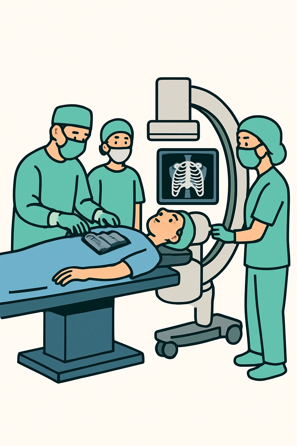
Intraoperative X-ray imaging is an X-ray taken during surgery. A portable X-ray machine or a C-arm device brings imaging into the operating room so the team can see bones, joints, or devices in real time. Surgeons use these images to guide steps in a procedure and to confirm placement and alignment before finishing. This can help reduce the need for repeat surgery and improve accuracy.
Also known as: Intraoperative radiograph, Intraop X-ray, Portable OR X-ray, Operative X-ray
Preparation & Next Steps
Everything you need to know before and after your procedure
Before Care
- Follow the surgery team’s fasting and medicine instructions; imaging is part of the procedure plan
- Bring prior imaging and reports or ensure the surgical team can access them to reduce repeat exposure
- Tell the team if you are or might be pregnant so shielding and timing can be considered
- Remove jewelry and removable metal near the operative area when asked, as metal can obscure images
- Share any history of reactions to iodine-based contrast, even though contrast is rarely used for plain X-rays
- Expect that a portable X-ray device or C-arm may be brought into the operating room with sterile covers
- Confirm who can receive updates during surgery and how results or images will be shared after
- Check your insurance benefits; imaging during surgery may appear as separate charges on the bill
After Care
- There is no separate recovery from the X-ray itself; recovery is determined by the surgery
- Radiation does not stay in the body after an X-ray
- Images are typically stored in the hospital system; you can request copies through the portal or medical records
- If a radiology report is created, review it in your portal when available
- Keep a personal list of imaging exams you have had to track cumulative exposure over time
- Expect that follow-up imaging may be ordered to check healing or device position
- Review your explanation of benefits; intraoperative imaging may be listed as a separate line item
- Contact the surgical team with any concerns about the imaging results or unexpected symptoms after surgery
Clinical Information
Important medical details about this procedure
Indications
- Confirming fracture reduction and alignment
- Checking placement of screws, plates, rods, or joint implants
- Verifying spinal level or hardware position
- Locating or confirming removal of a foreign body
- Guiding lines, tubes, or wires during procedures
- Counting or searching for retained surgical items when needed
Alternatives
- Preoperative or postoperative X-rays instead of imaging during surgery
- Fluoroscopy with live X-ray guidance
- Ultrasound guidance when suitable
- Intraoperative CT or cone-beam CT when available
- Optical or computer navigation systems using anatomic landmarks
Risks
- Small exposure to ionizing radiation to the patient
- Radiation exposure to operating room staff without proper protection
- Need for repeat images if the first images are unclear
- Rare allergic reaction if contrast is used for a related study
- Potential added time to the operation to obtain images
- Equipment positioning that may briefly disrupt workflow or sterility if not managed well
- Image misinterpretation that could affect decisions
Contraindications
- Known or possible pregnancy when imaging is not urgent
- History of severe contrast reaction if contrast would be used for a related study
- Extreme clinical instability where pausing for imaging would delay life-saving care
Recovery Timeline
What to expect during your recovery
Intraoperative X-ray imaging does not add recovery time on its own. Recovery depends on the underlying surgery.
Typical Range
Same day
Return to Work
Same day
Recovery Milestones
No additional recovery needs due to the imaging itself
Activity limits are determined by the surgery rather than the X-ray
Access images or the report in the patient portal if available
Frequently Asked Questions
Common questions and expert answers about this procedure
What is intraoperative X-ray imaging?
What is intraoperative X-ray imaging?
It is an X-ray taken during surgery using a portable machine or C-arm so the team can confirm alignment and device placement.
How is this different from fluoroscopy?
How is this different from fluoroscopy?
A radiograph is a still image taken at a moment in time. Fluoroscopy is live X-ray imaging. Some C-arm units can do both.
Does it hurt?
Does it hurt?
The X-ray itself is painless. You will be under anesthesia or sedation for the surgery, and positioning is managed by the team.
How much radiation is used?
How much radiation is used?
A single radiograph uses a small dose compared with CT. Dose varies by body part and equipment. Teams follow ALARA principles to minimize exposure.
Will there be a separate charge for the X-ray?
Will there be a separate charge for the X-ray?
Often yes. Charges can include the technical component for the equipment and a professional component if a radiologist interprets the image.
Are the images saved and can I see them?
Are the images saved and can I see them?
Images are usually stored in the hospital’s system. You can request copies or view them through the patient portal when available.
Is contrast used for intraoperative X-rays?
Is contrast used for intraoperative X-rays?
Plain radiographs do not use contrast. Some related procedures, such as arthrograms or angiography, may use contrast when needed.
How is staff and patient safety managed?
How is staff and patient safety managed?
The team uses shielding, limits the beam to the area of interest, and takes only the images needed to reduce exposure.
References
Medical literature and sources