%20--%3e%3csvg%20version='1.0'%20id='Layer_1'%20xmlns='http://www.w3.org/2000/svg'%20xmlns:xlink='http://www.w3.org/1999/xlink'%20x='0px'%20y='0px'%20viewBox='0%200%20755.52%20814.35'%20style='enable-background:new%200%200%20755.52%20814.35;'%20xml:space='preserve'%3e%3cstyle%20type='text/css'%3e%20.st0{fill:%231ae5ce;stroke:%231ae5ce;stroke-width:7;stroke-miterlimit:10;}%20.st1{fill:%23FFFFFF;stroke:%231ae5ce;stroke-width:7;stroke-miterlimit:10;}%20.st2{fill:%231ae5ce;stroke:%23FFFFFF;stroke-width:7;stroke-miterlimit:10;}%20.st3{fill:%23FFFFFF;stroke:%23FFFFFF;stroke-width:6;stroke-miterlimit:10;}%20%3c/style%3e%3cg%20id='Dark_Logo_00000159431215648494653180000015900878925932721032_'%3e%3cpath%20id='shield_00000152259091539869183100000001926543869452564382_'%20class='st0'%20d='M711.13,173.42%20c-3.37-49.07-38.92-44.9-75.96-52.18c-67.4-13.25-130.47-34.47-192.36-63.92c-22.48-10.7-41.88-25.74-65.05-26.28%20c-23.17,0.54-42.58,15.59-65.05,26.28c-61.89,29.45-124.95,50.68-192.36,63.92c-37.04,7.28-72.59,3.11-75.96,52.18%20c-1.55,22.6,0.34,46.4,1.05,68.92c5.99,190.78,52.91,345.39,206.27,466.46c29.71,23.46,74.51,55.8,108.84,70.75%20c5.99,2.61,11.7,3.73,17.21,3.75c5.51-0.02,11.22-1.14,17.21-3.75c34.33-14.95,79.12-47.3,108.84-70.75%20c153.37-121.07,200.28-275.68,206.27-466.46C710.79,219.82,712.68,196.02,711.13,173.42z'/%3e%3cpath%20id='magnifyingglass_00000073685934697636024790000000342567780940956808_'%20class='st1'%20d='M648.78,624.21l-96.64-104.65%20c34.89-40.63,55.98-93.46,55.98-151.21c0-128.27-103.98-232.24-232.24-232.24S143.63,240.08,143.63,368.35%20s103.98,232.24,232.24,232.24c48.87,0,94.21-15.11,131.62-40.89l97.18,105.24c11.25,12.18,30.24,12.93,42.42,1.69l0,0%20C659.27,655.38,660.03,636.39,648.78,624.21z'/%3e%3ccircle%20id='ellipse_00000129904010550392394720000017056154833629780114_'%20class='st2'%20cx='375.87'%20cy='368.35'%20r='181.21'/%3e%3cpath%20id='dollarsign_00000057140127156649725250000003772530148495155891_'%20class='st3'%20d='M362.91,492.03v-23.91%20c0-2.01-1.58-3.69-3.59-3.77c-18.49-0.78-37.12-5.4-49.4-11.83c-1.75-0.92-2.53-3.01-1.76-4.84l5.87-13.89%20c0.86-2.02,3.23-2.88,5.2-1.89c12.94,6.52,30.45,11.44,49.18,11.44c26.02,0,43.6-12.73,43.6-30.43c0-17.08-14.29-27.63-41.4-36.95%20c-37.37-12.42-60.46-26.7-60.46-53.72c0-24.97,20.29-44.11,52.22-49.51c1.8-0.31,3.11-1.89,3.11-3.72v-24.35%20c0-2.09,1.69-3.78,3.78-3.78h15.16c2.09,0,3.78,1.69,3.78,3.78v22.71c0,2.01,1.58,3.67,3.58,3.77c17.85,0.92,30.88,4.75,40.61,9.1%20c1.92,0.86,2.79,3.09,1.94,5.01l-6.08,13.6c-0.85,1.91-3.09,2.77-4.99,1.91c-8.37-3.8-22.38-8.95-42.75-8.95%20c-28.21,0-38.84,14.28-38.84,26.7c0,16.15,13.56,24.22,45.44,35.4c37.74,13.04,56.79,29.19,56.79,56.82%20c0,23.85-19.05,46.23-54.78,52.3c-1.8,0.31-3.11,1.89-3.11,3.72v25.28c0,2.09-1.69,3.78-3.78,3.78h-15.53%20C364.6,495.81,362.91,494.12,362.91,492.03z'/%3e%3c/g%3e%3c/svg%3e)
Knee X-ray (3 views) Diagnostic Imaging
A knee X-ray with 3 views is a set of three pictures taken with low-dose radiation to show the bones and joint space of the knee.
Overview
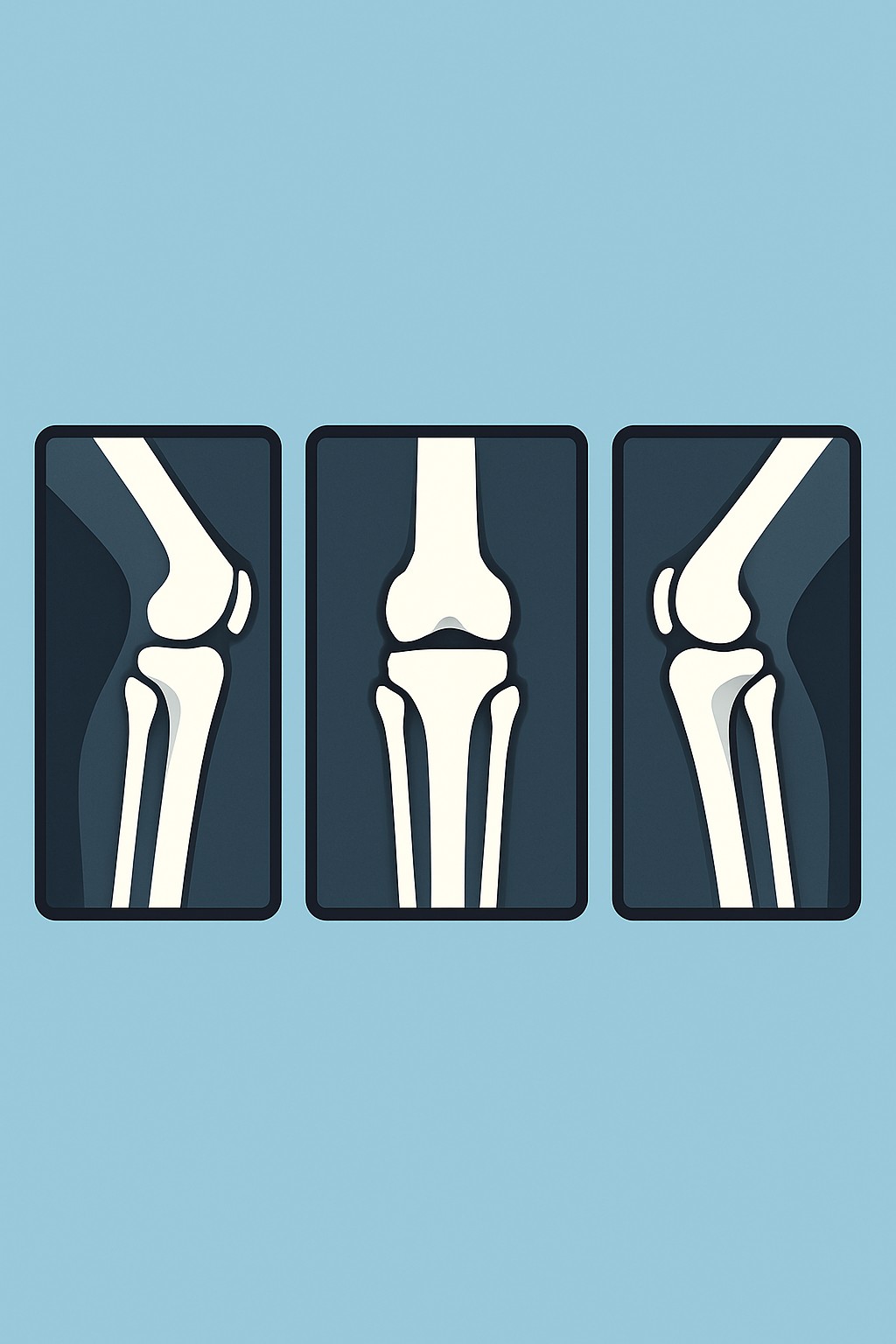
A knee X-ray with 3 views is a set of three pictures taken with low-dose radiation to show the bones and joint space of the knee. Common views include front (AP), side (lateral), and a kneecap view. These images help check alignment, joint spacing, and signs of injury or disease. Clinicians order a 3-view knee X-ray to look for fractures, arthritis changes, dislocations, or bone problems after pain, swelling, or trauma. It is quick, noninvasive, and often the first imaging test for knee concerns.
Also known as: Knee radiograph, Knee X-ray series, Knee 3-view X-ray
Preparation & Next Steps
Everything you need to know before and after your procedure
Before Care
- Bring your imaging order, photo ID, and any prior knee images or reports if available
- Wear comfortable clothing without metal around the knee; you may be asked to change into a gown
- Remove items that can block the image, such as jewelry, braces with metal, or bulky knee supports
- Tell the technologist if you could be pregnant or recently became pregnant
- Share brief history such as where the pain is, how the injury happened, and any prior knee surgery or hardware
- Arrive a bit early for check-in and to confirm the correct knee and side are imaged
- Use your usual mobility aids and ask for help if walking or standing is difficult
- Follow the facility’s instructions about patient portals if you want access to images or the report
- Ask about shielding if you have concerns about radiation exposure
After Care
- You can usually resume normal activities right after the X-ray
- Keep any splints, wraps, or supports applied by your clinic in place as instructed by your care team
- Check your patient portal for the radiology report when it is available, or ask how results will be shared
- Schedule follow-up with the ordering clinician to discuss findings and next steps
- Keep a copy of your images or know how to request them if needed for future visits
- If pain, swelling, or trouble moving the knee increases after the visit, contact a clinician
- Seek guidance from your care team if you receive new instructions based on the X-ray results
- Let the imaging center know if you notice any errors on your report, such as the wrong side labeled
Clinical Information
Important medical details about this procedure
Indications
- New knee pain after a fall or injury
- Suspected fracture or dislocation
- Swelling, limited motion, or instability
- Osteoarthritis evaluation (joint space narrowing, bone spurs)
- Suspected bone infection or tumor
- Checking healing or alignment after surgery or casting
- Evaluation of knee hardware position
Alternatives
- Clinical observation with rest, ice, compression, and elevation
- Ultrasound to look for fluid around the knee or some tendon issues
- MRI to assess ligaments, menisci, cartilage, and bone marrow
- CT scan for complex fractures or detailed bone evaluation
- Physical therapy and monitoring when imaging is not urgent
Risks
- Small exposure to ionizing radiation
- Incidental findings that may need further tests
- Discomfort from positioning the knee
- Need for repeat images if motion or clothing obscures the view
- Potential concerns during pregnancy if the abdomen is exposed
Contraindications
- Known or possible pregnancy without discussing risks and shielding
- Inability to remain still or safely position the knee
- Unstable trauma where immobilization is needed before imaging
Recovery Timeline
What to expect during your recovery
There is no medical recovery period from a standard knee X-ray. Most people return to normal activities right away.
Typical Range
Same day
Return to Work
Same day
Recovery Milestones
Resume normal daily activities
Review how results will be delivered (portal, phone, or follow-up)
Discuss results with the ordering clinician and confirm the plan
Frequently Asked Questions
Common questions and expert answers about this procedure
What does 3 views mean in a knee X-ray?
What does 3 views mean in a knee X-ray?
It is a set of three pictures of the knee from different angles, often front, side, and a view of the kneecap, to show more detail.
How long does a knee X-ray take?
How long does a knee X-ray take?
Imaging usually takes 10 to 15 minutes, though timing can vary with check-in and positioning.
Will a knee X-ray show ligament or meniscus tears?
Will a knee X-ray show ligament or meniscus tears?
X-rays show bones and joint spaces well. Ligaments and menisci are soft tissues and are better seen with MRI.
Is there radiation exposure?
Is there radiation exposure?
Yes, there is a small amount of ionizing radiation. For most people, the dose from a knee X-ray is low.
Can I get a knee X-ray if I am pregnant?
Can I get a knee X-ray if I am pregnant?
Tell the technologist if you are or might be pregnant. Imaging may be adjusted or delayed depending on need; shielding is often used.
Do I need to fast or stop medicines?
Do I need to fast or stop medicines?
No fasting is needed, and routine medicines are usually not affected for a standard knee X-ray.
Will it hurt?
Will it hurt?
The test itself does not hurt, but positioning may be uncomfortable if your knee is sore. Tell the technologist if a position is too painful.
When will I get results?
When will I get results?
A radiologist reviews the images and sends a report to the ordering clinician. Many centers post results in the patient portal.
References
Medical literature and sources