Leg vein ultrasound (bilateral) Diagnostic Imaging
A bilateral leg vein ultrasound is an imaging test that uses sound waves to look at the veins in both legs.
Overview
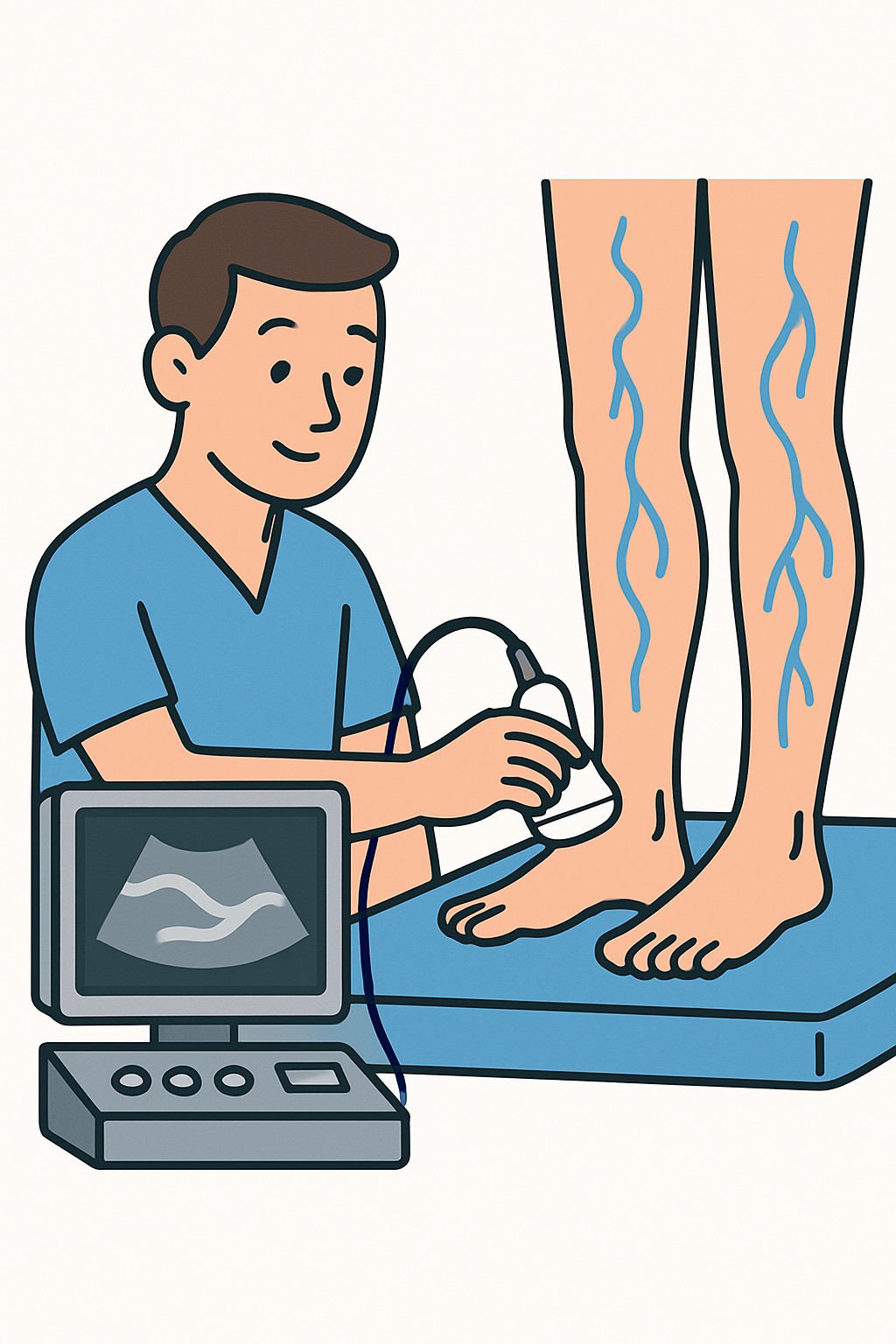
A bilateral leg vein ultrasound is an imaging test that uses sound waves to look at the veins in both legs. It shows how blood flows and whether the veins compress and open normally. Clinicians use this test to check for blood clots called deep vein thrombosis, to evaluate varicose veins and valve problems, and to map veins before or after vein procedures. It does not use radiation or contrast dye.
Also known as: Lower extremity venous duplex, Bilateral leg venous ultrasound, DVT ultrasound
Preparation & Next Steps
Everything you need to know before and after your procedure
Before Care
- Bring a photo ID, insurance card, and the imaging order or referral if provided
- Wear loose shorts or clothing that allows easy access from groin to ankle
- Avoid heavy lotions or oils on your legs the day of the exam so the gel makes good contact
- Confirm with the clinic whether to remove or pause compression stockings before arrival
- Arrive a bit early for check-in and to review any recent symptoms or surgeries
- Hydrate and use the restroom before the exam to stay comfortable during positioning
- Plan for 30 to 60 minutes, longer if detailed vein mapping is needed
- Bring a list of your medicines and any prior imaging or clot history
- If standing is difficult, tell the staff on arrival so they can adjust positioning
After Care
- Wipe off remaining gel and resume normal activities
- Confirm how and when results will be shared, such as through a patient portal or your clinician
- If additional imaging was suggested, note any scheduling instructions provided by the facility
- Keep a simple record of any ongoing leg symptoms to compare with results
- Contact a clinician if you notice new or worsening leg swelling, pain, redness, or shortness of breath
- Store the visit summary and any report copies in your personal health records
- Verify insurance coverage if follow-up tests or procedures are recommended
Clinical Information
Important medical details about this procedure
Indications
- Leg swelling, pain, warmth, or redness
- Suspected deep vein thrombosis
- Varicose veins and suspected venous insufficiency
- Skin changes on the lower legs such as darkening or sores
- Follow-up after a known blood clot or vein treatment
- Preoperative vein mapping for bypass or dialysis access
Alternatives
- Clinical risk assessment with blood tests such as D-dimer
- Repeat ultrasound at a later time if the first test is inconclusive
- CT venography
- MR venography
Risks
- Temporary discomfort from probe pressure on a tender area
- Skin irritation from ultrasound gel in rare cases
- False negative or false positive results that may require repeat testing
- Incidental findings that may lead to additional tests
Contraindications
- Open wounds, severe skin infection, or bulky dressings over the area
- Inability to remain still or follow positioning instructions
- Significant pain or limited mobility that prevents the exam positions
Recovery Timeline
What to expect during your recovery
There is no medical recovery period after a leg vein ultrasound. Most people return to normal activities right away.
Typical Range
Same day
Return to Work
Same day
Recovery Milestones
Resume normal daily activities
Review how your results will be delivered and check your portal
Complete any follow-up imaging or appointments if ordered
Frequently Asked Questions
Common questions and expert answers about this procedure
What does bilateral mean in this test?
What does bilateral mean in this test?
Bilateral means both legs are examined during the same visit.
How long does a bilateral leg vein ultrasound take?
How long does a bilateral leg vein ultrasound take?
Most exams take 30 to 60 minutes. Detailed vein mapping or reflux testing can take longer.
What conditions can this test detect?
What conditions can this test detect?
It can detect deep vein thrombosis, vein valve problems called venous insufficiency, and other flow issues.
Does it use radiation or contrast dye?
Does it use radiation or contrast dye?
No. Ultrasound uses sound waves. There is no ionizing radiation and no contrast dye.
Will the exam be painful?
Will the exam be painful?
It is usually painless. You may feel pressure from the probe, especially over a tender area.
Who performs and interprets the test?
Who performs and interprets the test?
A trained sonographer performs the scan. A radiologist or vascular specialist reviews the images and writes a report.
Do I need special preparation?
Do I need special preparation?
Wear loose clothing and avoid heavy lotions on your legs. The clinic will advise about compression stockings.
When will I get results?
When will I get results?
Timing varies by facility. Many centers send the report to your ordering clinician the same day or within a few days.
References
Medical literature and sources