Lumbar Spine X-Ray (2-3 views) Diagnostic Imaging
A lumbar spine X-ray takes pictures of the lower back using a small amount of ionizing radiation.
Overview
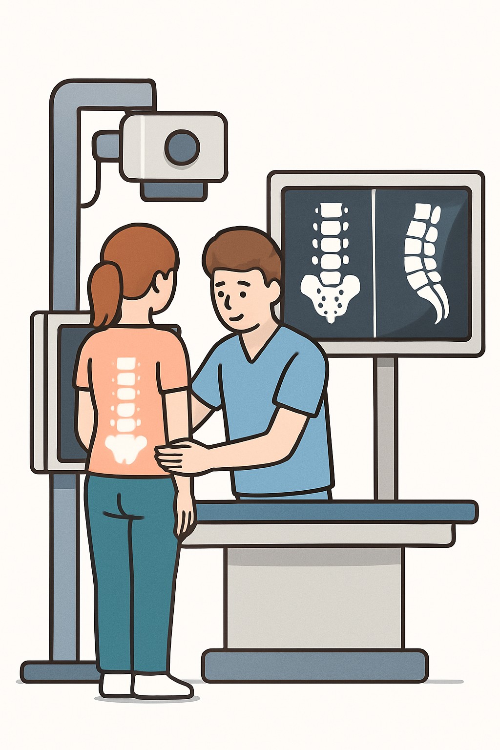
A lumbar spine X-ray takes pictures of the lower back using a small amount of ionizing radiation. The technologist usually captures 2 to 3 views from different angles, such as front, side, and sometimes oblique. Clinicians use these images to look at the bones of the lower back, including alignment, fractures, arthritis, and hardware from prior surgery. It does not show nerves, discs, or muscles as clearly as MRI or CT.
Also known as: Lumbar radiograph, Lumbar spine radiography, L-spine X-ray
Preparation & Next Steps
Everything you need to know before and after your procedure
Before Care
- Wear comfortable clothing without metal; you may be asked to change into a gown
- Remove jewelry, belts, keys, or anything with metal near the lower back
- Tell the technologist if you are or might be pregnant
- Bring prior spine images and reports if available for comparison
- Arrive a little early to allow time for check-in and safety screening
- Tell staff if you have severe pain, limited mobility, or cannot stand so positioning can be adjusted
- Let the team know about prior back surgery or implanted hardware
- No fasting or medication changes are typically needed unless the facility instructs otherwise
After Care
- Resume normal activities right after the X-ray
- Review your portal or the report when it becomes available
- Ask how and when results will be communicated and if follow-up imaging is expected
- Keep copies of your imaging reports for your records
- Tell your clinician if symptoms change, such as increasing pain, new numbness, or weakness
- If you might be pregnant and had an X-ray, inform your clinician so they can document exposure
Clinical Information
Important medical details about this procedure
Indications
- New or worsening low back pain with concerning features (such as recent trauma)
- Suspected fracture after a fall or accident
- Evaluation of spinal alignment or scoliosis
- Monitoring degenerative changes or arthritis
- Follow-up of known conditions, such as spondylolisthesis
- Check position and integrity of surgical hardware
Alternatives
- No immediate imaging for uncomplicated low back pain
- MRI to evaluate discs, nerves, and soft tissues
- CT for detailed bone evaluation
- Bone scan in select cases for stress injuries or tumors
- Physical exam and observation with conservative care
Risks
- Exposure to a small dose of ionizing radiation
- Incidental findings that may lead to more tests
- Discomfort from holding still in certain positions
- Limited detail for discs, nerves, and soft tissues, which could delay diagnosis if only an X-ray is obtained
- Special considerations if pregnant
Contraindications
- Known or possible pregnancy when imaging is not urgent
- Inability to remain still or follow positioning instructions without support
Recovery Timeline
What to expect during your recovery
There is no medical recovery period for a standard lumbar spine X-ray. Most people return to routine activities immediately.
Typical Range
Same day
Return to Work
Same day
Recovery Milestones
Resume normal daily activities
Access the imaging report in the patient portal if available
Discuss results and next steps with your clinician if recommended
Frequently Asked Questions
Common questions and expert answers about this procedure
What are 2–3 views in a lumbar spine X-ray?
What are 2–3 views in a lumbar spine X-ray?
They are pictures from different angles, commonly front (AP), side (lateral), and sometimes an oblique view to better see the joints and alignment.
How long does the exam take?
How long does the exam take?
The imaging itself usually takes about 10–15 minutes, though your total visit time can be longer due to check-in and positioning.
Do I need to fast or stop medicines?
Do I need to fast or stop medicines?
No special preparation is typically required. You can usually eat, drink, and take medicines as usual unless the facility gives different instructions.
Will it hurt?
Will it hurt?
The X-ray is painless. Some positions may be uncomfortable for a short time if you have back pain.
How much radiation is involved?
How much radiation is involved?
A lumbar spine X-ray uses a small dose of radiation. Typical effective dose is around 1–2 mSv, roughly equal to several months of natural background radiation.
Can I have this test if I am pregnant?
Can I have this test if I am pregnant?
X-rays are usually avoided in pregnancy unless clearly needed. Shielding and alternative imaging may be considered. Tell the staff if you are or might be pregnant.
What can a lumbar X-ray show and not show?
What can a lumbar X-ray show and not show?
It shows bones, alignment, fractures, arthritis, and hardware. It does not show nerves or discs well; MRI is better for those.
When are X-rays used for low back pain?
When are X-rays used for low back pain?
They are often used after trauma, with certain warning signs, or for persistent symptoms. For simple new low back pain, imaging may not be needed right away.
References
Medical literature and sources