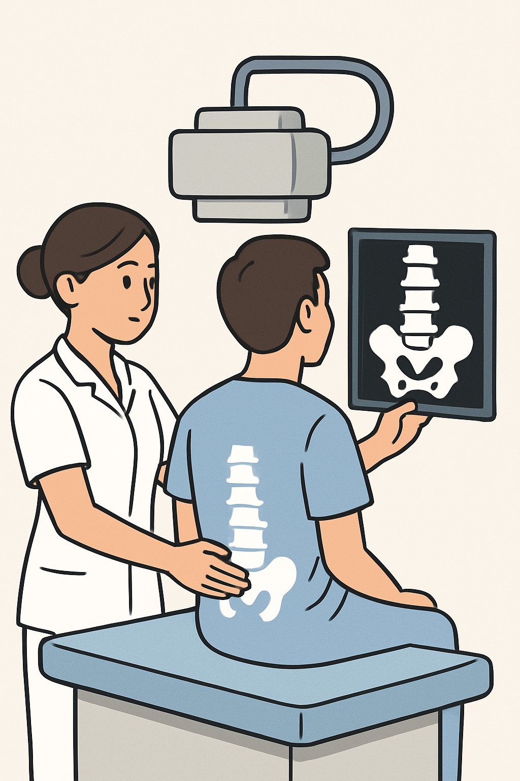
Lumbar spine X-ray (lower back) Diagnostic Imaging
A lumbar spine X-ray is an imaging test that uses a small dose of ionizing radiation to create pictures of the bones in the lower back.
Overview
A lumbar spine X-ray is an imaging test that uses a small dose of ionizing radiation to create pictures of the bones in the lower back. It shows the vertebrae, alignment, and visible changes in the joints and discs. Clinicians use it to look for fractures, arthritis, alignment problems, and changes after injury or surgery. It does not show nerves, muscles, or discs as clearly as MRI.
Also known as: Lower back X-ray, Lumbar X-ray, LS spine X-ray
Preparation & Next Steps
Everything you need to know before and after your procedure
Before Care
- Bring your imaging order and any prior spine images or reports if available
- Tell the imaging staff if you are or might be pregnant
- Wear comfortable clothing without metal zippers, snaps, or belts if possible
- Remove jewelry, wallets, and other metal objects from the waist and lower back area
- Arrive a little early to confirm identification, insurance, and consent forms
- Share any recent back injuries, surgeries, or implanted hardware with the technologist
- Ask how and when results will be delivered to your clinician
- No fasting or contrast is typically needed for a lumbar X-ray
After Care
- You can usually resume normal activities right away
- Confirm how you will receive results, such as through a patient portal or from your clinician
- Keep copies of images and reports for your records if offered
- If positioning caused temporary soreness, gentle movement and routine comfort measures may help
- Note any new or changing symptoms to discuss at follow-up
- Contact your clinician about concerning symptoms such as worsening back pain, new weakness, numbness, or changes in bowel or bladder control
- Ask whether additional imaging like MRI or CT is planned if questions remain after the X-ray
- Store any provided image CD or link in a safe place for future visits
Clinical Information
Important medical details about this procedure
Indications
- Lower back pain or stiffness
- Injury or trauma to the lower back
- Suspected fracture or compression fracture
- Evaluation of arthritis or degenerative changes
- Scoliosis or spondylolisthesis assessment
- Monitoring after spine surgery or hardware placement
- Unexplained back deformity or limited movement
Alternatives
- Observation and clinical follow-up
- Physical therapy and exercise programs
- MRI of the lumbar spine (better for nerves, discs, soft tissues)
- CT scan of the lumbar spine (detailed bone imaging)
- Bone scan when stress fracture or widespread bone issues are suspected
- Ultrasound for some soft tissue concerns near the back (limited use)
Risks
- Exposure to a small dose of ionizing radiation
- Potential risk to an unborn baby if pregnant
- Discomfort from positioning during the exam
- Limited ability to show soft tissues, which can lead to further testing
- Incidental findings that may need more evaluation
Contraindications
- Known or suspected pregnancy unless benefits outweigh risks
- Inability to remain still or follow positioning instructions
Recovery Timeline
What to expect during your recovery
There is no medical recovery period after a standard lumbar spine X-ray. Most people return to normal activities immediately.
Typical Range
Same day
Return to Work
Same day
Recovery Milestones
Resume normal daily activities
Review how results will be communicated and plan follow-up if needed
Discuss next steps with your clinician if symptoms persist or change
Frequently Asked Questions
Common questions and expert answers about this procedure
What does a lumbar spine X-ray show?
What does a lumbar spine X-ray show?
It shows bones in the lower back, including alignment, fractures, and arthritis. It does not clearly show nerves, discs, muscles, or ligaments.
How long does the exam take?
How long does the exam take?
The imaging itself usually takes about 10 to 15 minutes. Time can vary with positioning and clinic workflow.
Will it hurt?
Will it hurt?
The X-ray is painless. Some people feel brief discomfort from holding certain positions.
Is there radiation exposure?
Is there radiation exposure?
Yes, it uses a small dose of ionizing radiation. Facilities aim to use the lowest dose that produces a clear image.
Is it safe during pregnancy?
Is it safe during pregnancy?
X-rays are generally avoided in pregnancy when possible. If imaging is needed, shielding and alternative tests may be considered.
Do I need to fast or get contrast?
Do I need to fast or get contrast?
No. A standard lumbar X-ray does not use contrast and does not require fasting.
How is it different from MRI or CT?
How is it different from MRI or CT?
X-rays show bones and alignment well. MRI shows nerves and discs better. CT shows detailed bone structures and some soft tissue.
When will I get results?
When will I get results?
A radiologist reviews the images and sends a report to your clinician. Timing varies by facility and may appear in your patient portal.
References
Medical literature and sources