MRI Lumbar Spine (without/with contrast) Diagnostic Imaging
An MRI of the lumbar spine uses a strong magnet and radio waves to create detailed pictures of the lower back.
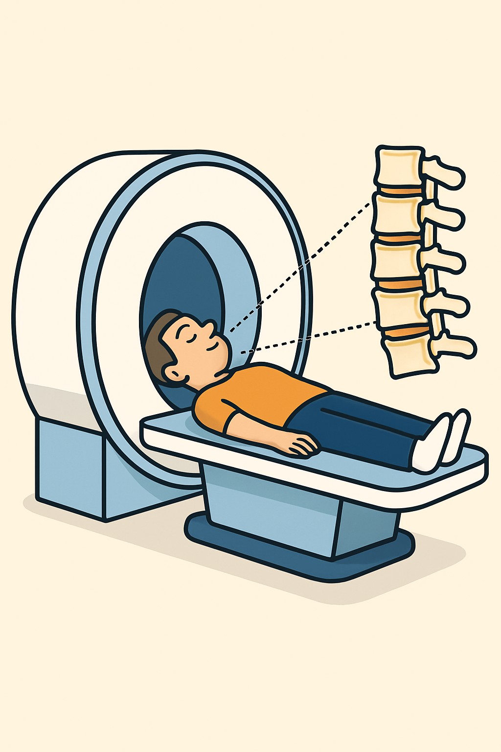
Overview
An MRI of the lumbar spine uses a strong magnet and radio waves to create detailed pictures of the lower back. The images show bones, discs, nerves, muscles, and other soft tissues without using radiation. Without/with contrast means the scan starts without contrast dye and then continues after a small amount of gadolinium contrast is given through an IV, if needed. Contrast can make certain tissues and blood vessels easier to see and can help tell old from new changes.
Also known as: Lumbar spine MRI, MRI lower back, MRI lumbar without and with contrast
Preparation & Next Steps
Everything you need to know before and after your procedure
Before Care
- Complete the MRI safety screening and share details of any implants, surgeries, or metal fragments, especially in the eyes.
- Bring device cards or make and model information for pacemakers, stimulators, pumps, clips, or joint hardware.
- Remove jewelry, piercings, hairpins, watches, and other metal items; wear clothing without zippers or snaps if possible.
- Confirm any recent imaging and bring prior spine images or reports to help with comparison.
- Tell the staff about kidney problems, prior contrast reactions, asthma, or allergies; a recent kidney function test may be requested when contrast is planned.
- Discuss claustrophobia or anxiety ahead of time; ask about an open scanner, music, or calming medicine if the site offers it.
- Follow the imaging center’s instructions about eating and drinking, especially if contrast or sedation may be used.
- Arrange a ride if you expect to receive a sedative; some centers require an escort for discharge.
- Arrive early to allow time for check-in, IV placement, and screening.
- Stay hydrated unless you were told otherwise.
After Care
- Most people resume normal activities right after the scan.
- If contrast was used, drink fluids through the day unless you were told otherwise.
- Watch the IV site for redness, swelling, warmth, or pain.
- If you notice hives, itching, wheezing, or trouble breathing soon after the exam, contact the imaging center or your clinician.
- Follow the center’s instructions if you were given a sedative, including transportation and activity limits.
- Ask how and when your results will be available; many centers release reports and images through a patient portal.
- Keep a copy of your images and report for your records or future consultations.
- Share any implanted device information with your care team for future imaging planning.
- Let your clinician know if pain, numbness, or weakness changes after the scan.
- Schedule any recommended follow-up visits or tests as directed by your care team.
Clinical Information
Important medical details about this procedure
Indications
- Low back pain that does not improve or has red flags
- Leg pain, numbness, or weakness suggesting nerve compression (sciatica)
- Suspected disc herniation or spinal stenosis
- Known or suspected cancer affecting the spine
- Suspected infection in the spine or discs
- Evaluation of fractures or other injuries not fully seen on other imaging
- Preoperative planning or postoperative assessment
- Suspected inflammatory or demyelinating conditions affecting the lower spine
- Evaluation for cauda equina symptoms such as severe weakness or bladder changes
Alternatives
- MRI without contrast alone when adequate
- X-ray of the lumbar spine
- CT scan of the lumbar spine
- CT myelography when MRI is not possible
- Electrodiagnostic testing (EMG/NCS) for nerve function
- Watchful waiting with follow-up
- Physical therapy and home exercise programs
- Medicines and activity modification
Risks
- Feeling confined or anxious in the scanner
- Loud sounds that may be uncomfortable without hearing protection
- Discomfort from lying still
- Heating or movement of some metal implants or fragments; screening reduces this risk
- IV contrast risks such as nausea, headache, or rare allergic reactions
- Gadolinium contrast is generally avoided in severe kidney disease due to risk of a rare condition called nephrogenic systemic fibrosis
- Small amounts of gadolinium may remain in the body; health effects are not clearly known
- Bruising or soreness at the IV site
Contraindications
- Certain pacemakers, defibrillators, or implants that are not MRI-conditional
- Some aneurysm clips, cochlear implants, or metal fragments in or near the eyes
- Severe kidney failure for the use of gadolinium contrast
- History of severe reaction to gadolinium-based contrast
- Inability to lie still or severe claustrophobia without support options
- Use of gadolinium contrast during pregnancy is generally avoided unless clearly needed
Recovery Timeline
What to expect during your recovery
There is usually no medical recovery period after an MRI of the lumbar spine. People typically return to normal activities the same day. A short observation period may be needed if sedation was used.
Typical Range
Same day
Return to Work
Same day
Recovery Milestones
Resume normal daily activities
Increase fluids if contrast was used, unless instructed otherwise
Monitor IV site for redness or swelling
Review imaging report and plan follow-up with your care team
Frequently Asked Questions
Common questions and expert answers about this procedure
What does without/with contrast mean for a lumbar MRI?
What does without/with contrast mean for a lumbar MRI?
Images are taken first without contrast and then again after a small amount of gadolinium contrast is given through an IV. Contrast can make inflammation, tumors, blood vessels, and postoperative changes easier to see.
How long does the scan take?
How long does the scan take?
Most lumbar spine MRIs take 30 to 60 minutes. Adding contrast can extend the time by about 10 to 20 minutes.
Does an MRI use radiation?
Does an MRI use radiation?
No. MRI uses a strong magnet and radio waves. It does not use ionizing radiation.
Can I get this MRI if I have a pacemaker or other implant?
Can I get this MRI if I have a pacemaker or other implant?
Some devices are MRI-conditional and can be scanned with special steps. Others are not safe. The imaging team checks device type and settings before scheduling.
Is gadolinium contrast safe for kidneys?
Is gadolinium contrast safe for kidneys?
In people with severe kidney problems, some contrast agents have been linked to a rare condition called nephrogenic systemic fibrosis. Centers screen for kidney disease and choose contrast carefully.
What if I am claustrophobic?
What if I am claustrophobic?
Options may include an open or wider scanner, music or a mirror, or a short-acting calming medicine. Tell the center in advance so they can plan.
Can I breastfeed after receiving gadolinium contrast?
Can I breastfeed after receiving gadolinium contrast?
Professional groups state breastfeeding can continue after gadolinium. If you have concerns, discuss options with your care team before the scan.
How will I get my results?
How will I get my results?
A radiologist reads the images and sends a report to your ordering clinician. Many centers also release the report and images to your patient portal.
References
Medical literature and sources