Obstetric Ultrasound (Prenatal) Diagnostic Imaging
An obstetric ultrasound is an imaging test that uses sound waves to create pictures of a pregnancy.
Overview
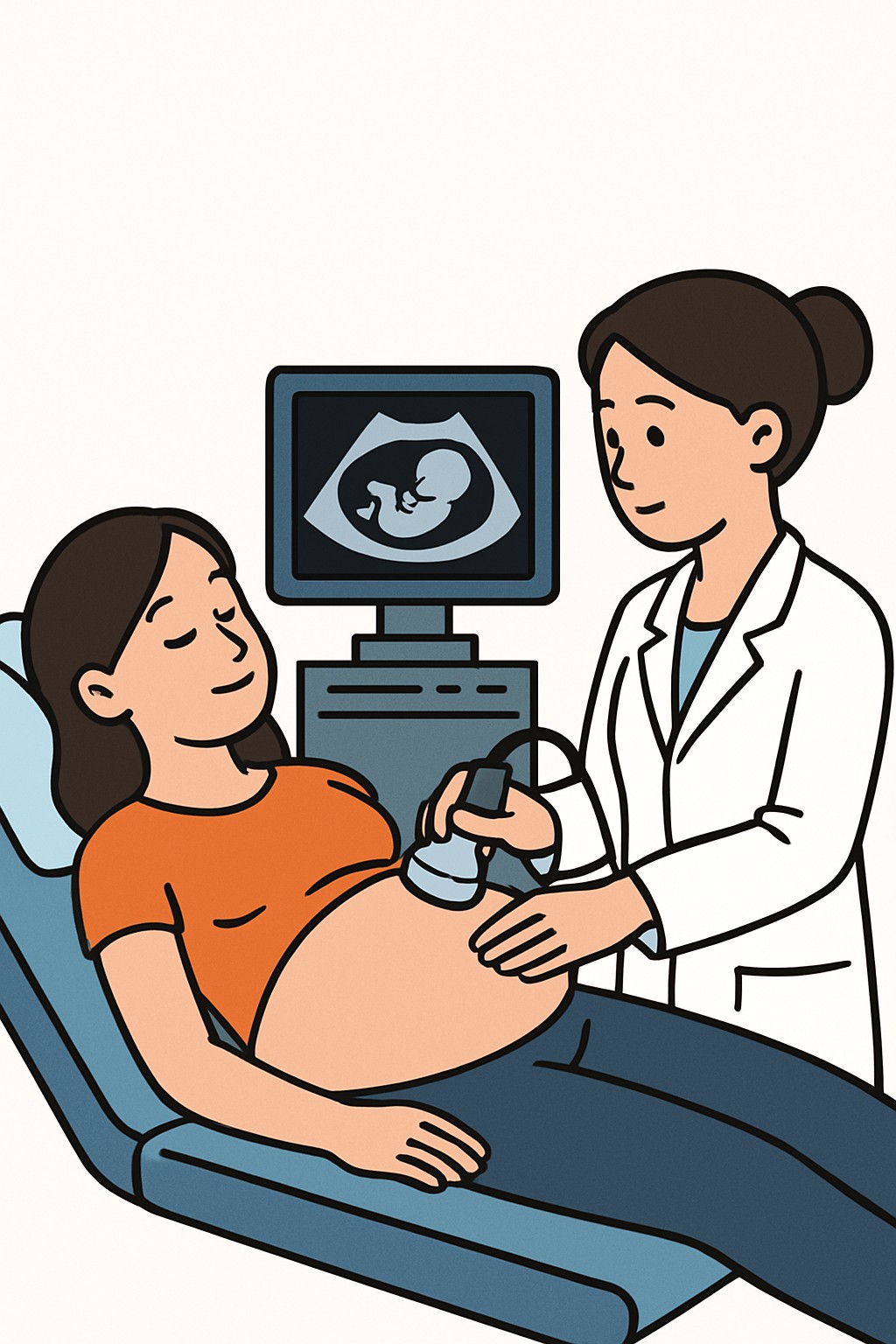
An obstetric ultrasound is an imaging test that uses sound waves to create pictures of a pregnancy. It does not use ionizing radiation. A clinician or sonographer moves a small handheld device on the abdomen, or uses a thin internal probe in the vagina early in pregnancy, to see the uterus, placenta, and fetus. This test helps check gestational age, number of fetuses, fetal heartbeat, growth, anatomy, amniotic fluid, and placental location. It can guide care plans and may help find problems that need closer follow-up.
Also known as: Prenatal ultrasound, Fetal ultrasound, OB ultrasound, Pregnancy ultrasound
Preparation & Next Steps
Everything you need to know before and after your procedure
Before Care
- Bring a government ID and any referral or order if provided by your clinician
- Wear a two-piece outfit so the abdomen is easy to access
- Some centers ask for a comfortably full bladder for early scans; follow the facility instructions
- You may be offered a transvaginal exam early in pregnancy for clearer images; ask about this option
- Confirm the site policy on guests and whether images will be provided
- Arrive a few minutes early to update contact information and medical history
- Tell staff about any allergies to latex or ultrasound gel
- Bring prior imaging reports or prenatal records if available
- Ask how long the appointment is expected to take and plan transportation as needed
After Care
- You can return to normal activities right after the exam
- Wipe off any remaining gel and check clothing for residue
- Light spotting can occur after a transvaginal exam; contact your clinic if you have heavy bleeding, severe pain, or fever
- Review the final report in your patient portal once it is released
- Write down any questions about the findings to discuss with your care team
- If a follow-up scan was suggested, note the timing window so scheduling stays on track
- Keep any printed or digital images with your personal records if they were provided
- Report new or worsening symptoms to your care team
Clinical Information
Important medical details about this procedure
Indications
- Confirming pregnancy location and dating early in pregnancy
- Checking fetal heartbeat and number of fetuses
- Anatomy survey in mid pregnancy
- Monitoring fetal growth and amniotic fluid
- Assessing placenta position or bleeding
- Evaluating pain, cramping, or other concerning symptoms
- Following up abnormal screening or prior imaging
- Guiding procedures such as amniocentesis
Alternatives
- Clinical exam and fundal height measurements
- Maternal blood tests and genetic screening
- Fetal MRI in selected situations
- Observation with repeat clinical assessment
- Auscultation of fetal heart with stethoscope or fetoscope when feasible
Risks
- Generally considered safe when used appropriately by trained professionals
- No ionizing radiation; theoretical heating or mechanical effects if used excessively
- Mild discomfort from probe pressure or a full bladder
- Transvaginal exam may feel invasive and can be followed by light spotting
- Incidental or uncertain findings that may require more tests
- Nonmedical keepsake ultrasounds are discouraged by regulators
Contraindications
- No common absolute contraindications when medically indicated
- Latex sensitivity to probe covers; latex-free options may be needed
- Recent pelvic surgery or active infection may limit transvaginal approach
- Avoid nonmedical keepsake scans that are not ordered for care
Recovery Timeline
What to expect during your recovery
Most people resume normal activities immediately after an obstetric ultrasound. There is no medical recovery period.
Typical Range
Same day
Return to Work
Same day
Recovery Milestones
Resume normal daily activities
If transvaginal exam was done, expect possible light spotting that typically resolves
Review final report in portal and note any recommended follow-up imaging
Frequently Asked Questions
Common questions and expert answers about this procedure
What is the difference between transabdominal and transvaginal ultrasound?
What is the difference between transabdominal and transvaginal ultrasound?
Transabdominal scans use a probe on the belly with gel. Transvaginal scans use a thin probe placed a short distance into the vagina for closer views early in pregnancy.
Is obstetric ultrasound safe for the baby?
Is obstetric ultrasound safe for the baby?
Ultrasound uses sound waves, not X-rays. When used appropriately by trained professionals, it is considered safe. Regulators advise against nonmedical keepsake scans.
When are ultrasounds commonly done in pregnancy?
When are ultrasounds commonly done in pregnancy?
An early scan may confirm dating and location. A detailed anatomy scan is commonly performed in the middle of pregnancy based on the care plan.
Do I need a full bladder?
Do I need a full bladder?
Some centers ask for a comfortably full bladder early in pregnancy to improve images. Instructions vary by facility.
How long does the exam take?
How long does the exam take?
Most exams take about 15 to 60 minutes, depending on gestational age, fetal position, and how detailed the study is.
Will I get results right away?
Will I get results right away?
You may hear general impressions during the visit. A final report is typically reviewed by a specialist and then shared with you, often in the patient portal.
Can I get 3D or 4D images?
Can I get 3D or 4D images?
3D or 4D imaging may be used when medically useful. Routine keepsake imaging without a medical reason is discouraged by the FDA.
What happens if something unusual is seen?
What happens if something unusual is seen?
Your care team may suggest repeat or targeted ultrasound, lab tests, or referral to a specialist. Next steps depend on the specific finding.
References
Medical literature and sources