Post-void bladder residual ultrasound Diagnostic Imaging
A post-void bladder residual ultrasound is a noninvasive test that estimates how much urine remains in the bladder after you urinate.
Overview
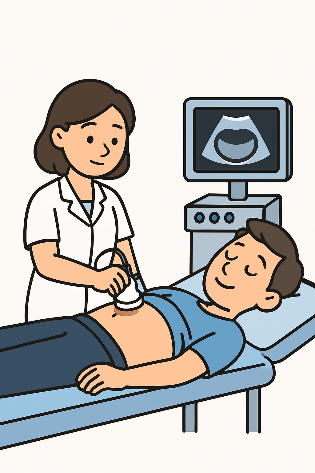
A post-void bladder residual ultrasound is a noninvasive test that estimates how much urine remains in the bladder after you urinate. A small ultrasound probe is placed on the lower belly with gel, and the machine calculates the bladder volume using sound waves. Clinicians use this test to see how well the bladder empties. It can help evaluate symptoms such as weak stream, hesitancy, or leakage and can guide next steps in care.
Also known as: PVR ultrasound, Post-void residual scan, Bladder scan after voiding, Bladder ultrasound PVR
Preparation & Next Steps
Everything you need to know before and after your procedure
Before Care
- Confirm any clinic instructions about arriving with a full bladder or voiding on arrival
- Drink water ahead of time only if instructed; avoid emptying your bladder until staff directs you
- Wear two-piece clothing to allow easy access to the lower abdomen
- Bring a current list of medicines, especially diuretics, and any recent urinary test results
- Note the time you last urinated before the appointment
- Share any mobility or continence needs so staff can plan restroom access
- Plan extra time in case repeat scans are needed after additional voiding
- If you use intermittent catheterization, ask the clinic how to handle it around the scan
- Most centers do not require fasting; verify any special preparation when scheduling
After Care
- Wipe off remaining gel; it does not stain and should wash away easily
- Resume usual activities and fluid intake unless your clinician provided different instructions
- Review results in your patient portal or visit summary when available
- If more testing was discussed, schedule appointments and note any preparation steps
- Track urinary symptoms in a simple diary to support future visits
- Contact the clinic if you develop fever, severe lower abdominal pain, inability to urinate, or worsening urinary symptoms
- Ask how results will be communicated if you do not see them within the expected timeframe
- Keep any follow-up appointments to review findings and next steps
Clinical Information
Important medical details about this procedure
Indications
- Urinary retention or difficulty emptying
- Weak urine stream or hesitancy
- Feeling of incomplete bladder emptying
- Overflow incontinence or nighttime leakage
- Recurrent urinary tract infections
- Evaluation of benign prostatic hyperplasia (BPH)
- Neurogenic bladder or nerve conditions affecting urination
- Monitoring after pelvic or urinary surgery
- Pelvic organ prolapse symptoms
- Assessment after urinary catheter removal
Alternatives
- Measuring residual urine with a straight catheter (catheterization)
- Bedside portable bladder scanner instead of formal imaging
- Uroflowmetry to measure urine flow pattern
- Bladder diary and timed voiding as supportive assessments
Risks
- Mild discomfort from a full bladder or probe pressure
- Cold sensation or temporary skin irritation from gel
- Inaccurate result if the timing after urination is off or technique is suboptimal
- Privacy or modesty concerns during the exam
Contraindications
- No known absolute contraindications to diagnostic ultrasound
- Open wounds, dressings, or severe skin irritation over the lower abdomen
- Situations needing urgent catheterization for acute urinary retention
- Limited ability to follow instructions for voiding and positioning
Recovery Timeline
What to expect during your recovery
This is a noninvasive imaging test with no medical recovery period. Most people return to normal activities immediately after the scan.
Typical Range
Same day
Return to Work
Same day
Recovery Milestones
Resume normal daily activities
Review results when posted or communicated
Complete any follow-up tests if they were ordered
Frequently Asked Questions
Common questions and expert answers about this procedure
What is a post-void residual?
What is a post-void residual?
It is the amount of urine left in the bladder right after you urinate. The ultrasound estimates this volume without using a catheter.
How is the test done?
How is the test done?
You are asked to urinate, then lie back while a clinician places an ultrasound probe with gel over the lower belly to estimate the remaining bladder volume.
How long does it take?
How long does it take?
The scan itself usually takes a few minutes. The visit can take longer if repeat measurements are needed.
Is there any radiation?
Is there any radiation?
No. Ultrasound uses sound waves, not ionizing radiation.
Will it hurt?
Will it hurt?
Most people feel only mild pressure from the probe and the cool gel. Discomfort is uncommon.
Do I need special preparation?
Do I need special preparation?
Fasting is not typical. Clinics may ask you to arrive with a comfortably full bladder or to wait to void until the scan is set up.
How accurate is it?
How accurate is it?
Results depend on timing after urination and proper technique. If a very precise measurement is needed, catheterization may be used.
What can the results suggest?
What can the results suggest?
A higher residual can be seen with blockage, weak bladder muscle, or nerve problems. A low residual suggests better bladder emptying.
References
Medical literature and sources