Rib cage X-ray (multiple views) Diagnostic Imaging
A rib cage X-ray uses a small dose of radiation to create pictures of the ribs and chest wall.
Overview
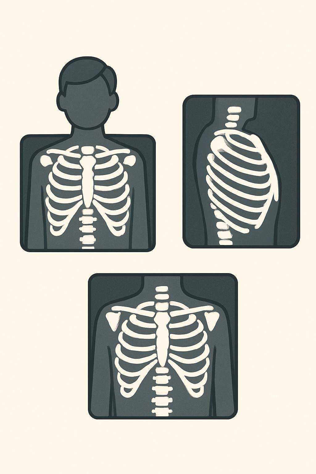
A rib cage X-ray uses a small dose of radiation to create pictures of the ribs and chest wall. Multiple views means the technologist takes images from different angles, often with you standing or lying in varied positions. You may be asked to hold your breath briefly during each image. This test is commonly used to look for broken ribs, bone changes, or signs of injury to the chest wall. It can also help check for complications related to rib injuries, such as problems in the area near the lungs.
Also known as: Rib series, Rib X-ray, Ribs radiograph, Chest wall X-ray
Preparation & Next Steps
Everything you need to know before and after your procedure
Before Care
- Bring your photo ID, insurance information, and any imaging order or referral if required
- Wear loose, comfortable clothing; avoid metal snaps or graphics over the chest; remove necklaces and piercings
- Tell the imaging staff if you are or might be pregnant so they can plan shielding or consider alternatives
- Bring prior rib or chest images or reports, or know where they are available in your patient portal
- Arrive a bit early to check in and complete forms
- Be prepared to hold your breath and change positions for multiple views
- Let staff know about pain, limited movement, or breathing issues so they can position you safely
- No fasting is usually needed for a rib X-ray
- If you use mobility aids or oxygen, bring them and any settings information
- Confirm how and when results will be communicated and who will receive the report
After Care
- You can usually return to normal activities right after the X-ray
- There is no radiation left in your body after the exam
- Check how and when results will be available from your ordering clinician or patient portal
- Keep track of your imaging history if you monitor radiation exposure
- Follow any plans for additional imaging or evaluation if requested by your clinician
- Contact a clinician if pain, swelling, or trouble breathing worsens after the injury
- Make sure any new imaging reports are shared with the clinician managing your care
- Bring questions about the findings to your next visit or send a secure message to the clinic
Clinical Information
Important medical details about this procedure
Indications
- Rib pain or tenderness after a fall, crash, or sports injury
- Suspected rib fracture
- Chest pain that worsens with deep breaths or movement
- Localized chest wall swelling or bruising
- Follow-up on a known rib fracture to assess healing
- Evaluation of possible bone lesions or abnormalities in the ribs
- Chest wall deformity or suspected congenital rib differences
Alternatives
- Chest X-ray to evaluate lungs and pleura when rib injury is suspected
- Ultrasound to look for rib fractures or fluid around the lung
- CT scan of the chest for detailed bone and lung images
- MRI for soft tissue or bone marrow changes
- Bone scan for stress fractures or widespread bone issues
- Observation and clinical follow-up without immediate imaging
Risks
- Exposure to a small amount of ionizing radiation
- Limited ability to show very small or non-displaced fractures
- Discomfort from positioning or holding breath during images
- Incidental findings that may need more testing
- Need for additional imaging if views are unclear
Contraindications
- Known or possible pregnancy without discussing protective measures with imaging staff
- Inability to stay still or follow breath-hold instructions
- Severe pain or injury that prevents safe positioning
- Objects or clothing with metal over the chest that cannot be removed
Recovery Timeline
What to expect during your recovery
There is no medical recovery period from a rib X-ray. Most people resume normal activity immediately.
Typical Range
Same day
Return to Work
Same day
Recovery Milestones
Resume normal daily activities
Access results in the portal or from the ordering clinician
Review results and confirm any follow-up imaging or visit
Frequently Asked Questions
Common questions and expert answers about this procedure
What does multiple views mean?
What does multiple views mean?
The technologist takes images from different angles and positions to see the ribs clearly. You may stand, sit, or lie down and briefly hold your breath.
How long does a rib X-ray take?
How long does a rib X-ray take?
The imaging itself usually takes a few minutes. Positioning and check-in can make the total visit about 15 to 30 minutes.
Will the X-ray show a small fracture?
Will the X-ray show a small fracture?
It can show many fractures, but tiny or non-displaced fractures may not be visible. If concern remains, other imaging like ultrasound or CT may be considered.
Is there radiation exposure?
Is there radiation exposure?
Yes, a small amount. X-rays use ionizing radiation, but doses for diagnostic rib images are typically kept as low as reasonably achievable.
Can I have a rib X-ray if I am pregnant?
Can I have a rib X-ray if I am pregnant?
Imaging is sometimes performed with shielding and careful technique when the benefits outweigh the risks. Tell the staff if you are or might be pregnant.
Do I need contrast or an injection?
Do I need contrast or an injection?
No. Standard rib X-rays do not use contrast dye or injections.
What is the difference between a rib X-ray and a chest X-ray?
What is the difference between a rib X-ray and a chest X-ray?
A rib X-ray focuses on the bones of the chest wall. A chest X-ray is aimed at the lungs, heart, and nearby structures.
Will it hurt?
Will it hurt?
The X-ray is painless, but holding certain positions can be uncomfortable, especially after an injury. Tell the technologist if a position hurts.
References
Medical literature and sources