%20--%3e%3csvg%20version='1.0'%20id='Layer_1'%20xmlns='http://www.w3.org/2000/svg'%20xmlns:xlink='http://www.w3.org/1999/xlink'%20x='0px'%20y='0px'%20viewBox='0%200%20755.52%20814.35'%20style='enable-background:new%200%200%20755.52%20814.35;'%20xml:space='preserve'%3e%3cstyle%20type='text/css'%3e%20.st0{fill:%231ae5ce;stroke:%231ae5ce;stroke-width:7;stroke-miterlimit:10;}%20.st1{fill:%23FFFFFF;stroke:%231ae5ce;stroke-width:7;stroke-miterlimit:10;}%20.st2{fill:%231ae5ce;stroke:%23FFFFFF;stroke-width:7;stroke-miterlimit:10;}%20.st3{fill:%23FFFFFF;stroke:%23FFFFFF;stroke-width:6;stroke-miterlimit:10;}%20%3c/style%3e%3cg%20id='Dark_Logo_00000159431215648494653180000015900878925932721032_'%3e%3cpath%20id='shield_00000152259091539869183100000001926543869452564382_'%20class='st0'%20d='M711.13,173.42%20c-3.37-49.07-38.92-44.9-75.96-52.18c-67.4-13.25-130.47-34.47-192.36-63.92c-22.48-10.7-41.88-25.74-65.05-26.28%20c-23.17,0.54-42.58,15.59-65.05,26.28c-61.89,29.45-124.95,50.68-192.36,63.92c-37.04,7.28-72.59,3.11-75.96,52.18%20c-1.55,22.6,0.34,46.4,1.05,68.92c5.99,190.78,52.91,345.39,206.27,466.46c29.71,23.46,74.51,55.8,108.84,70.75%20c5.99,2.61,11.7,3.73,17.21,3.75c5.51-0.02,11.22-1.14,17.21-3.75c34.33-14.95,79.12-47.3,108.84-70.75%20c153.37-121.07,200.28-275.68,206.27-466.46C710.79,219.82,712.68,196.02,711.13,173.42z'/%3e%3cpath%20id='magnifyingglass_00000073685934697636024790000000342567780940956808_'%20class='st1'%20d='M648.78,624.21l-96.64-104.65%20c34.89-40.63,55.98-93.46,55.98-151.21c0-128.27-103.98-232.24-232.24-232.24S143.63,240.08,143.63,368.35%20s103.98,232.24,232.24,232.24c48.87,0,94.21-15.11,131.62-40.89l97.18,105.24c11.25,12.18,30.24,12.93,42.42,1.69l0,0%20C659.27,655.38,660.03,636.39,648.78,624.21z'/%3e%3ccircle%20id='ellipse_00000129904010550392394720000017056154833629780114_'%20class='st2'%20cx='375.87'%20cy='368.35'%20r='181.21'/%3e%3cpath%20id='dollarsign_00000057140127156649725250000003772530148495155891_'%20class='st3'%20d='M362.91,492.03v-23.91%20c0-2.01-1.58-3.69-3.59-3.77c-18.49-0.78-37.12-5.4-49.4-11.83c-1.75-0.92-2.53-3.01-1.76-4.84l5.87-13.89%20c0.86-2.02,3.23-2.88,5.2-1.89c12.94,6.52,30.45,11.44,49.18,11.44c26.02,0,43.6-12.73,43.6-30.43c0-17.08-14.29-27.63-41.4-36.95%20c-37.37-12.42-60.46-26.7-60.46-53.72c0-24.97,20.29-44.11,52.22-49.51c1.8-0.31,3.11-1.89,3.11-3.72v-24.35%20c0-2.09,1.69-3.78,3.78-3.78h15.16c2.09,0,3.78,1.69,3.78,3.78v22.71c0,2.01,1.58,3.67,3.58,3.77c17.85,0.92,30.88,4.75,40.61,9.1%20c1.92,0.86,2.79,3.09,1.94,5.01l-6.08,13.6c-0.85,1.91-3.09,2.77-4.99,1.91c-8.37-3.8-22.38-8.95-42.75-8.95%20c-28.21,0-38.84,14.28-38.84,26.7c0,16.15,13.56,24.22,45.44,35.4c37.74,13.04,56.79,29.19,56.79,56.82%20c0,23.85-19.05,46.23-54.78,52.3c-1.8,0.31-3.11,1.89-3.11,3.72v25.28c0,2.09-1.69,3.78-3.78,3.78h-15.53%20C364.6,495.81,362.91,494.12,362.91,492.03z'/%3e%3c/g%3e%3c/svg%3e)
Spine X-ray (lumbosacral 2-3 views) Diagnostic Imaging
A lumbosacral spine X-ray uses a small amount of radiation to create pictures of the bones in the lower back (lumbar spine) and sacrum.
Overview
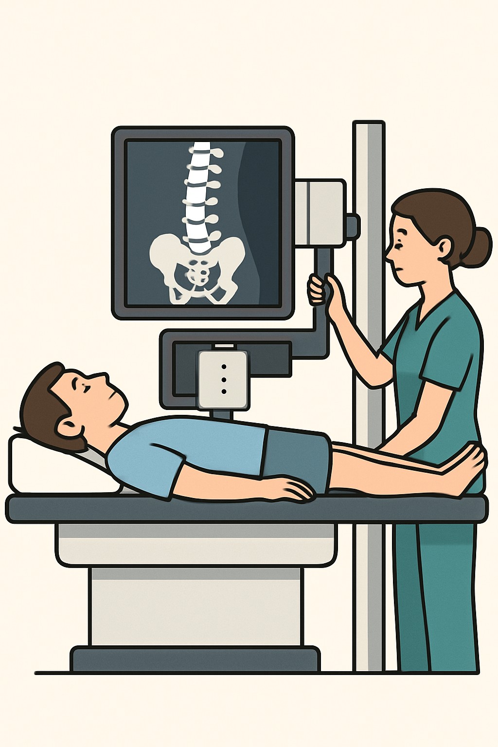
A lumbosacral spine X-ray uses a small amount of radiation to create pictures of the bones in the lower back (lumbar spine) and sacrum. The technologist takes 2 to 3 views, usually from the front, from the side, and sometimes a focused view of the L5–S1 level, to show alignment and bone detail. This test is used to look for fractures, alignment problems, arthritis changes, or other bone issues. It does not show soft tissues like discs, nerves, or muscles as clearly as MRI or CT.
Also known as: Lumbar spine X-ray, Lumbosacral spine radiograph, LS spine X-ray, Lumbar X-ray
Preparation & Next Steps
Everything you need to know before and after your procedure
Before Care
- Bring the imaging order, photo ID, insurance card, and any prior spine images or reports for comparison
- Wear comfortable clothing without metal zippers, snaps, or buttons; remove belts, jewelry, and pocket items
- Inform the staff if there is any chance of pregnancy
- Let the technologist know about prior spine surgery, metal implants, or a back brace
- No fasting is usually required; follow any facility-specific instructions provided at scheduling
- Arrive a bit early to complete registration and safety screening
- Ask about assistance if standing or lying flat is difficult due to pain or mobility limits
- Silence phones and secure valuables before entering the imaging room
- Confirm how and when results will be delivered (portal, report to ordering clinician)
After Care
- Resume normal activities right after the X-ray
- Any stiffness from holding a position usually passes quickly
- Radiation does not remain in the body after an X-ray
- A radiologist reviews the images and sends a report to the ordering clinician
- Check your patient portal or with the clinic to learn when the report is available
- Keep copies of images or reports if provided for your records
- Contact a clinician if symptoms change significantly or new concerning symptoms appear
- Additional imaging such as MRI or CT may be suggested based on the X-ray findings and your symptoms
Clinical Information
Important medical details about this procedure
Indications
- Lower back pain after an injury or fall
- Suspected fracture or spine alignment problem
- Known or suspected spondylolisthesis (slipped vertebra)
- Degenerative changes such as osteoarthritis
- Scoliosis or other curvature assessment
- Follow-up of prior surgery or hardware position
- Monitoring known bone conditions of the lumbar spine
Alternatives
- Observation and physical examination
- MRI for discs, nerves, or soft tissues
- CT scan for detailed bone evaluation
- Bone scan for stress injury or widespread bone changes
- Physical therapy and symptom tracking when immediate imaging is not required
Risks
- Low dose radiation exposure to the lower back and pelvis
- Potential risk to an unborn baby if performed during pregnancy
- Discomfort from holding still or certain positions
- Limited ability to show soft-tissue problems such as disc herniation
- Incidental findings that may lead to additional tests
Contraindications
- Known or possible pregnancy unless benefits outweigh risks
- Inability to remain still or safely position due to severe pain or injury
- Unstable trauma where spine motion must be minimized until stabilized
Recovery Timeline
What to expect during your recovery
There is no medical recovery period after a lumbosacral spine X-ray. Most people return to usual activities immediately.
Typical Range
Same day
Return to Work
Same day
Recovery Milestones
Resume normal daily activities
Review the portal or clinic messages for the radiology report
Discuss results and next steps with the ordering clinician if planned
Frequently Asked Questions
Common questions and expert answers about this procedure
What does 2 to 3 views mean?
What does 2 to 3 views mean?
It refers to taking pictures from different angles, usually front and side, and sometimes a focused view of the L5–S1 level for added detail.
How long does a lumbosacral spine X-ray take?
How long does a lumbosacral spine X-ray take?
The exam itself usually takes about 10 to 15 minutes, depending on positioning and the number of views.
Will this show a herniated disc or pinched nerve?
Will this show a herniated disc or pinched nerve?
X-rays show bones well but not soft tissues like discs or nerves. MRI is often used to evaluate those structures.
Is the radiation exposure high?
Is the radiation exposure high?
The dose is small. It is higher than a chest X-ray and lower than many CT scans. Facilities use the lowest dose needed for clear images.
Can I have this test if I am pregnant?
Can I have this test if I am pregnant?
Imaging that uses X-rays is generally avoided during pregnancy unless clearly needed. Tell the technologist if pregnancy is possible.
Do I need an injection or contrast dye?
Do I need an injection or contrast dye?
No. A standard lumbosacral spine X-ray uses external X-rays and does not use contrast.
Is it safe with metal implants or a back brace?
Is it safe with metal implants or a back brace?
Metal can appear on the images but is not harmful. You may be asked to remove a removable brace or external metal for clearer pictures.
How will I get the results?
How will I get the results?
A radiologist interprets the images and sends a report to the ordering clinician. Results are often available in the portal or from the clinic within a short time.
References
Medical literature and sources