%20--%3e%3csvg%20version='1.0'%20id='Layer_1'%20xmlns='http://www.w3.org/2000/svg'%20xmlns:xlink='http://www.w3.org/1999/xlink'%20x='0px'%20y='0px'%20viewBox='0%200%20755.52%20814.35'%20style='enable-background:new%200%200%20755.52%20814.35;'%20xml:space='preserve'%3e%3cstyle%20type='text/css'%3e%20.st0{fill:%231ae5ce;stroke:%231ae5ce;stroke-width:7;stroke-miterlimit:10;}%20.st1{fill:%23FFFFFF;stroke:%231ae5ce;stroke-width:7;stroke-miterlimit:10;}%20.st2{fill:%231ae5ce;stroke:%23FFFFFF;stroke-width:7;stroke-miterlimit:10;}%20.st3{fill:%23FFFFFF;stroke:%23FFFFFF;stroke-width:6;stroke-miterlimit:10;}%20%3c/style%3e%3cg%20id='Dark_Logo_00000159431215648494653180000015900878925932721032_'%3e%3cpath%20id='shield_00000152259091539869183100000001926543869452564382_'%20class='st0'%20d='M711.13,173.42%20c-3.37-49.07-38.92-44.9-75.96-52.18c-67.4-13.25-130.47-34.47-192.36-63.92c-22.48-10.7-41.88-25.74-65.05-26.28%20c-23.17,0.54-42.58,15.59-65.05,26.28c-61.89,29.45-124.95,50.68-192.36,63.92c-37.04,7.28-72.59,3.11-75.96,52.18%20c-1.55,22.6,0.34,46.4,1.05,68.92c5.99,190.78,52.91,345.39,206.27,466.46c29.71,23.46,74.51,55.8,108.84,70.75%20c5.99,2.61,11.7,3.73,17.21,3.75c5.51-0.02,11.22-1.14,17.21-3.75c34.33-14.95,79.12-47.3,108.84-70.75%20c153.37-121.07,200.28-275.68,206.27-466.46C710.79,219.82,712.68,196.02,711.13,173.42z'/%3e%3cpath%20id='magnifyingglass_00000073685934697636024790000000342567780940956808_'%20class='st1'%20d='M648.78,624.21l-96.64-104.65%20c34.89-40.63,55.98-93.46,55.98-151.21c0-128.27-103.98-232.24-232.24-232.24S143.63,240.08,143.63,368.35%20s103.98,232.24,232.24,232.24c48.87,0,94.21-15.11,131.62-40.89l97.18,105.24c11.25,12.18,30.24,12.93,42.42,1.69l0,0%20C659.27,655.38,660.03,636.39,648.78,624.21z'/%3e%3ccircle%20id='ellipse_00000129904010550392394720000017056154833629780114_'%20class='st2'%20cx='375.87'%20cy='368.35'%20r='181.21'/%3e%3cpath%20id='dollarsign_00000057140127156649725250000003772530148495155891_'%20class='st3'%20d='M362.91,492.03v-23.91%20c0-2.01-1.58-3.69-3.59-3.77c-18.49-0.78-37.12-5.4-49.4-11.83c-1.75-0.92-2.53-3.01-1.76-4.84l5.87-13.89%20c0.86-2.02,3.23-2.88,5.2-1.89c12.94,6.52,30.45,11.44,49.18,11.44c26.02,0,43.6-12.73,43.6-30.43c0-17.08-14.29-27.63-41.4-36.95%20c-37.37-12.42-60.46-26.7-60.46-53.72c0-24.97,20.29-44.11,52.22-49.51c1.8-0.31,3.11-1.89,3.11-3.72v-24.35%20c0-2.09,1.69-3.78,3.78-3.78h15.16c2.09,0,3.78,1.69,3.78,3.78v22.71c0,2.01,1.58,3.67,3.58,3.77c17.85,0.92,30.88,4.75,40.61,9.1%20c1.92,0.86,2.79,3.09,1.94,5.01l-6.08,13.6c-0.85,1.91-3.09,2.77-4.99,1.91c-8.37-3.8-22.38-8.95-42.75-8.95%20c-28.21,0-38.84,14.28-38.84,26.7c0,16.15,13.56,24.22,45.44,35.4c37.74,13.04,56.79,29.19,56.79,56.82%20c0,23.85-19.05,46.23-54.78,52.3c-1.8,0.31-3.11,1.89-3.11,3.72v25.28c0,2.09-1.69,3.78-3.78,3.78h-15.53%20C364.6,495.81,362.91,494.12,362.91,492.03z'/%3e%3c/g%3e%3c/svg%3e)
Transvaginal Ultrasound (OB or Gyn) Diagnostic Imaging
A transvaginal ultrasound uses a small covered probe placed a short distance inside the vagina to create pictures of the uterus, cervix, ovaries, and nearby structures.
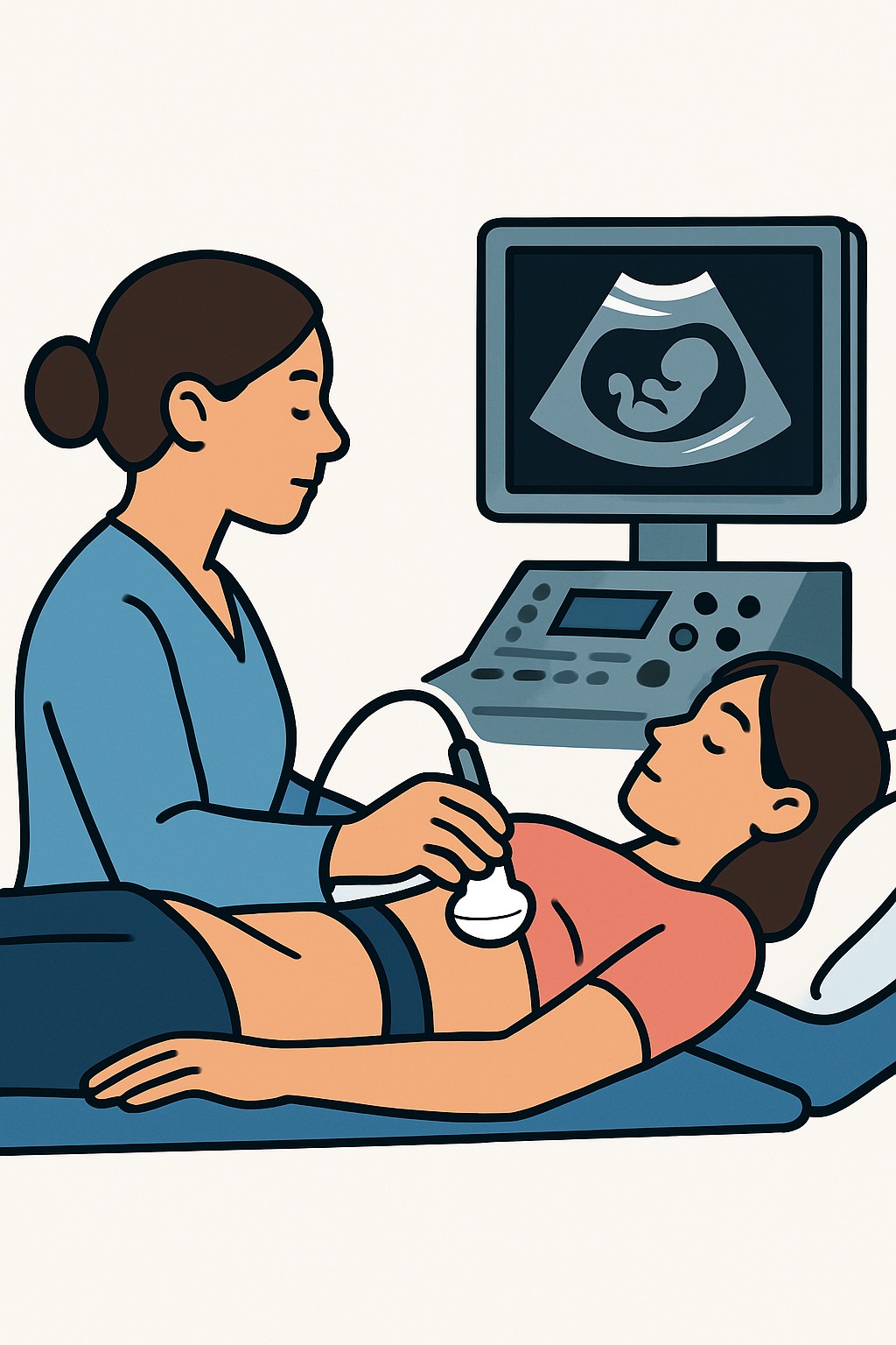
Overview
A transvaginal ultrasound uses a small covered probe placed a short distance inside the vagina to create pictures of the uterus, cervix, ovaries, and nearby structures. It uses sound waves, not radiation. In obstetrics, it is often used early in pregnancy to confirm location and dating, check for causes of bleeding, and assess the cervix. In gynecology, it helps evaluate pelvic pain, abnormal bleeding, infertility, ovarian cysts, fibroids, and the position of an intrauterine device.
Also known as: Endovaginal ultrasound, Pelvic ultrasound (transvaginal), Transvaginal sonogram, TVUS
Preparation & Next Steps
Everything you need to know before and after your procedure
Before Care
- Bring a photo ID, any imaging order, and your insurance card if you have one
- Know the date of your last menstrual period and current pregnancy status if applicable
- You may be asked to empty your bladder before the exam; follow the facility’s instructions
- Tell the staff about any latex allergy so a non-latex cover can be used
- Wear a two-piece outfit for easier changing
- Remove tampons or menstrual cups before the exam
- Bring a list of your medicines and any relevant prior imaging or reports
- Arrive a bit early to review consent and check-in forms
- Ask the clinic in advance about chaperone availability if desired
After Care
- You can return to normal activities right after the exam
- Mild cramping or light spotting can happen for a short time
- Use pads rather than internal products immediately after if spotting occurs
- Review how and when you will receive results, such as through a patient portal
- Schedule any follow-up imaging or appointments if recommended
- Keep copies of your report for future visits
- Contact a clinician if you develop heavy bleeding, fever, or severe pain
- If pregnant, note any new or worsening symptoms and inform your obstetric team
Clinical Information
Important medical details about this procedure
Indications
- Early pregnancy dating and viability
- Confirming intrauterine vs ectopic pregnancy
- Vaginal bleeding or pelvic pain in pregnancy
- Measuring cervical length during pregnancy
- Abnormal uterine bleeding
- Pelvic pain or pressure
- Evaluation of ovarian cysts, masses, or fibroids
- Infertility workup, including follicle monitoring
- Checking intrauterine device position
- Follow-up of known pelvic conditions
Alternatives
- Transabdominal pelvic ultrasound
- Pelvic MRI when detailed soft tissue imaging is needed
- Pelvic exam and laboratory tests based on symptoms
- CT pelvis in select urgent situations
- Watchful waiting with follow-up imaging when appropriate
Risks
- Temporary discomfort or pressure during probe insertion
- Light spotting may occur afterward
- Very small risk of infection
- Allergic reaction to latex if a latex probe cover is used
- Possibility of unclear images requiring repeat or different imaging
- Ultrasound uses no ionizing radiation
Contraindications
- No consent to an internal exam
- Severe pain or recent pelvic surgery that prevents probe insertion
- Latex allergy if a non-latex cover is not available
- Individuals who have not had vaginal intercourse and prefer external imaging
Recovery Timeline
What to expect during your recovery
Most people resume normal activities immediately after a transvaginal ultrasound. Any mild spotting or cramping usually resolves quickly.
Typical Range
Same day
Return to Work
Same day
Recovery Milestones
Resume normal daily activities
Expect mild spotting or cramping to resolve if present
Review report and arrange any follow-up recommended by your clinician
Frequently Asked Questions
Common questions and expert answers about this procedure
What happens during a transvaginal ultrasound?
What happens during a transvaginal ultrasound?
A covered, lubricated probe is placed a short distance into the vagina. The technologist or clinician gently moves it to capture images of the uterus, cervix, ovaries, and nearby areas.
Is it safe during pregnancy?
Is it safe during pregnancy?
Yes. Ultrasound uses sound waves, not ionizing radiation. It is commonly used in early pregnancy and to measure cervical length.
Do I need a full or empty bladder?
Do I need a full or empty bladder?
For transvaginal imaging, an empty bladder is often preferred. Follow the instructions from the facility.
Will it hurt?
Will it hurt?
Most people feel pressure and brief discomfort. Tell the staff if you are uncomfortable so they can adjust the exam.
How long does it take?
How long does it take?
The exam typically takes about 15 to 30 minutes, depending on the reason for the study and the views needed.
Can it be done during menstruation?
Can it be done during menstruation?
Yes. Imaging quality is usually not affected. You may be asked to remove tampons or menstrual cups before the exam.
Who performs and interprets the test?
Who performs and interprets the test?
A sonographer or clinician performs the scan. A radiologist or obstetric-gynecologic clinician interprets the images and issues a report.
References
Medical literature and sources