Ultrasound of chest (e.g. pleural fluid scan) Diagnostic Imaging
An ultrasound of the chest is a noninvasive imaging test that uses sound waves to create pictures of the tissues just inside the chest wall.
Overview
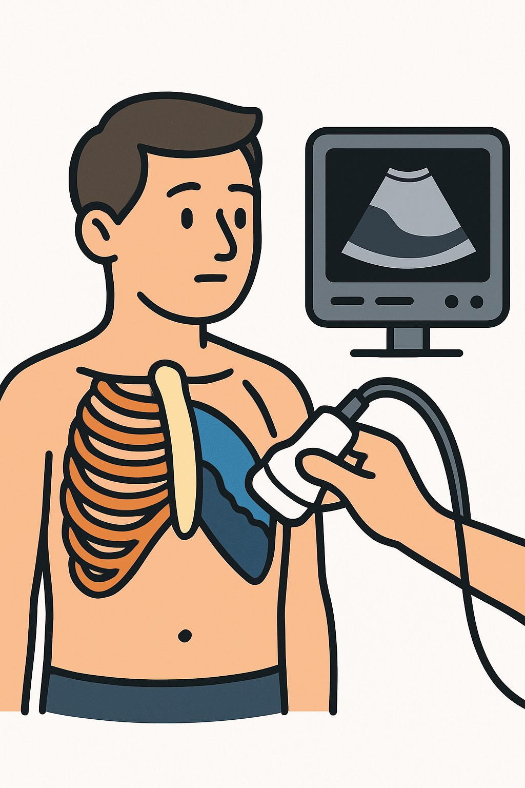
An ultrasound of the chest is a noninvasive imaging test that uses sound waves to create pictures of the tissues just inside the chest wall. It helps visualize the pleura (the lining around the lungs), pleural fluid, the diaphragm, and parts of the lung surface. Clinicians use chest ultrasound to look for pleural effusion (fluid around the lungs), signs that suggest pneumonia or lung collapse near the surface, and to check diaphragm movement. It is also commonly used to guide procedures such as thoracentesis, helping improve accuracy and reduce complications. Ultrasound does not use ionizing radiation.
Also known as: Chest ultrasound, Lung ultrasound, Pleural ultrasound, Thoracic ultrasound
Preparation & Next Steps
Everything you need to know before and after your procedure
Before Care
- No fasting is usually needed; confirm any special instructions if the scan will guide a procedure
- Wear a loose two-piece outfit; you may be asked to change into a gown
- Avoid heavy lotions, oils, or powders on the chest the day of the exam to help the probe make contact
- Bring a current list of medicines, allergies, and any prior reactions to gels or adhesives
- Carry prior chest imaging reports or discs if available for comparison
- Tell the team about breathing symptoms, recent injuries, surgeries, or chest drains
- Arrive a bit early to complete check-in and verify your preferred pharmacy and contact info
- Ask whether a support person is allowed if communication or mobility is a concern
- If imaging will guide thoracentesis or another procedure, expect additional prep from that care team
- Confirm how and when you will receive the results (portal, call, or follow-up visit)
After Care
- The technologist will wipe off the gel; you can typically resume normal activities right away
- A radiologist or trained clinician reviews the images and sends a report to the ordering clinician
- Check your patient portal or clinic messages for the report when available
- Keep track of symptoms such as shortness of breath, chest pain, or fever and share changes with your care team
- If ultrasound guided a procedure, follow the separate instructions you received for site care and activity
- Note any new redness, swelling, or drainage at a puncture site after a related procedure and contact a clinician if it occurs
- Keep copies of the report and images if offered for your records
- Ask about next steps if the scan shows fluid or other findings, such as monitoring, more imaging, or procedures
Clinical Information
Important medical details about this procedure
Indications
- Suspected or known pleural effusion
- Evaluation for pneumothorax near the chest wall
- Assessment of pneumonia or consolidation close to the pleura
- Guidance for thoracentesis or chest tube placement
- Monitoring known fluid collections over time
- Evaluation of chest wall masses or rib fractures
- Assessment of diaphragm movement or paralysis
- Shortness of breath with unclear cause after initial exam
Alternatives
- Chest X-ray
- CT scan of the chest
- MRI of the chest in selected situations
- Observation with repeat clinical exam
- Lung function tests when appropriate
Risks
- Mild discomfort from probe pressure or cool gel
- Temporary skin irritation or rare gel allergy
- Limited views in people with dressings, wounds, or body habitus
- False negatives or positives that may lead to further tests
- If used to guide a procedure, risks relate to that procedure rather than the ultrasound itself
Contraindications
- No common absolute contraindications
- Open wounds, burns, or bulky dressings over the scan area may limit imaging
- Severe pain or inability to remain still can reduce image quality
Recovery Timeline
What to expect during your recovery
Chest ultrasound does not require recovery time. Most people return to normal activities immediately after the exam.
Typical Range
Same day
Return to Work
Same day
Recovery Milestones
Resume normal daily activities
Review preliminary findings or the formal report when available
Complete any scheduled follow-up imaging or appointments
Frequently Asked Questions
Common questions and expert answers about this procedure
What is a chest ultrasound?
What is a chest ultrasound?
It is an imaging test that uses sound waves to create pictures of the pleura, pleural fluid, chest wall, diaphragm, and the outer parts of the lungs.
Does a chest ultrasound use radiation?
Does a chest ultrasound use radiation?
No. Ultrasound uses sound waves, not ionizing radiation.
What can a chest ultrasound detect?
What can a chest ultrasound detect?
It commonly detects pleural effusion, helps assess pneumothorax near the chest wall, shows some infections or collapse close to the pleura, and evaluates diaphragm movement.
How long does the exam take?
How long does the exam take?
Many scans take about 15 to 30 minutes, but timing varies by the reason for the exam and whether it guides a procedure.
Do I need to fast or stop medicines?
Do I need to fast or stop medicines?
Fasting is usually not needed. If the ultrasound will guide a procedure like thoracentesis, the care team may give extra instructions.
Who performs and interprets the test?
Who performs and interprets the test?
A sonographer or trained clinician performs the scan. A radiologist or qualified clinician interprets the images and sends a report.
Will the ultrasound hurt?
Will the ultrasound hurt?
It is generally painless. You may feel slight pressure from the probe or cool gel on the skin.
What happens if fluid is found?
What happens if fluid is found?
Options may include monitoring, additional imaging, or a procedure such as thoracentesis. The care team explains choices based on the overall findings.
References
Medical literature and sources