Visual Field Exam (Automated perimetry) Diagnostic Imaging
An automated visual field exam measures how wide and sensitive your field of vision is, especially your side (peripheral) vision.
Overview
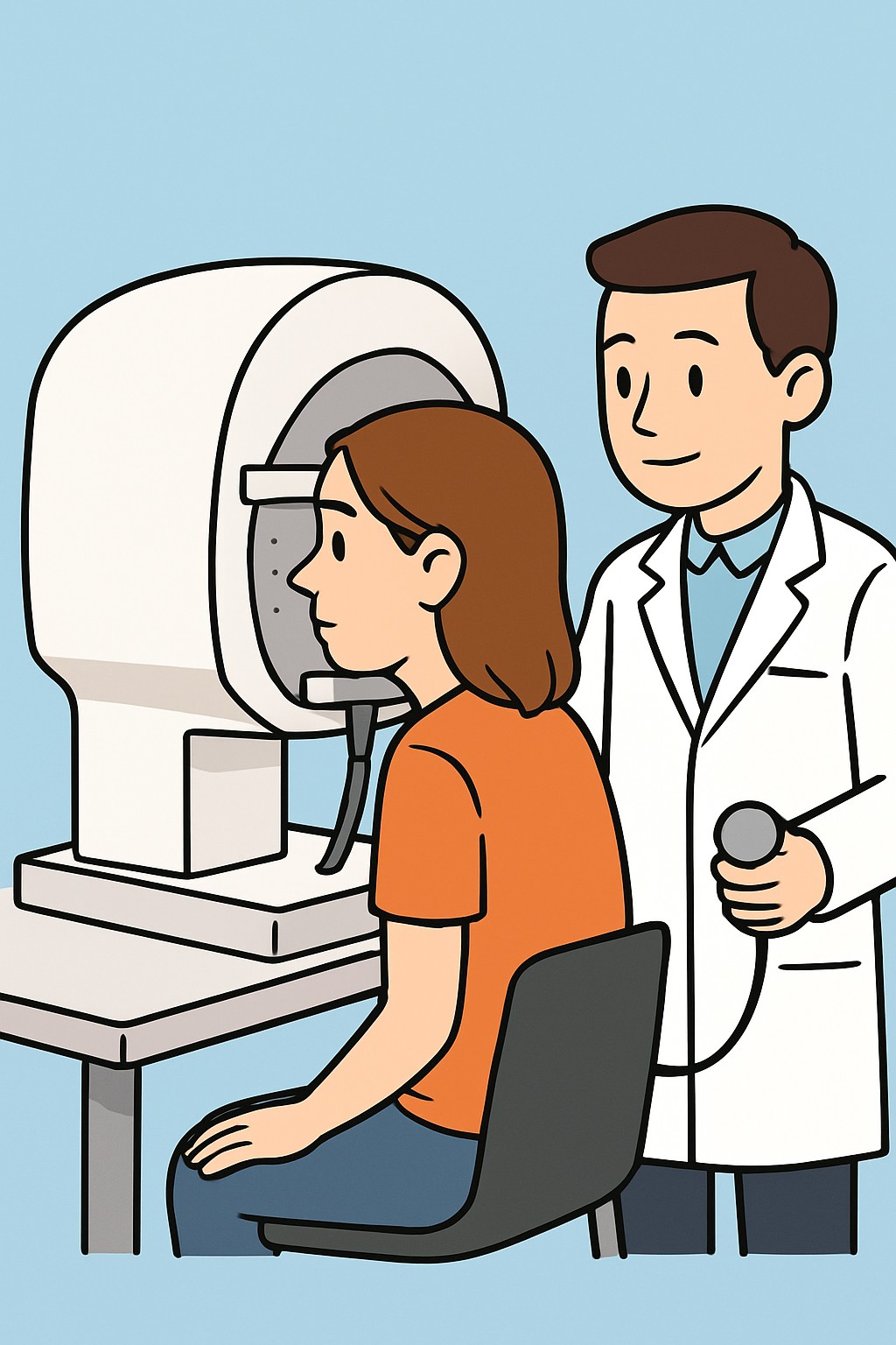
An automated visual field exam measures how wide and sensitive your field of vision is, especially your side (peripheral) vision. You look into a dome-shaped machine and press a button when you see small lights. The device maps any areas where vision may be reduced. Clinicians use this test to check for damage to the optic nerve or pathways from the eye to the brain. It is commonly used to diagnose and monitor glaucoma and other conditions that can affect peripheral vision.
Also known as: Automated visual field test, Perimetry, Humphrey visual field
Preparation & Next Steps
Everything you need to know before and after your procedure
Before Care
- Bring your current glasses and a copy of your most recent prescription if available
- Confirm with the clinic whether dilating eye drops will be used during the visit and plan for temporary light sensitivity
- Wear comfortable clothing and plan to sit still for 10–20 minutes per eye
- Arrive a bit early to review instructions on how to respond during the test
- Let staff know about any hearing, mobility, or communication needs so they can adjust the setup
- Share a list of your medicines, especially any that affect alertness or vision
- Rest beforehand and avoid scheduling the test when you are very tired to help with attention
- If dilation is planned and you are sensitive to light, bring sunglasses and consider arranging a ride
After Care
- You can resume normal activities right after the test
- If your pupils were dilated for the visit, expect light sensitivity and blurry near vision for several hours
- Use sunglasses in bright light if dilation was done
- Review when and how you will receive results and any follow-up plans
- Tell the clinic if you had trouble focusing, staying alert, or pressing the button, as this can affect results
- Keep any follow-up appointment to compare results over time
- Report new or worsening vision changes to your eye care team
Clinical Information
Important medical details about this procedure
Indications
- Diagnosing and monitoring glaucoma
- Evaluating unexplained vision loss or blind spots
- Assessing vision changes after stroke, brain tumor, or pituitary disease
- Monitoring optic nerve diseases such as optic neuritis
- Evaluating retinal conditions that affect peripheral vision
- Screening for medicine effects on the retina or optic nerve (for example, some long-term therapies)
Alternatives
- Confrontation visual field test at the bedside
- Manual perimetry (Goldmann kinetic perimetry)
- Targeted eye exam and imaging as clinically appropriate (for example, OCT), often used alongside perimetry
Risks
- Temporary eye strain or fatigue
- Mild headache or frustration from the repetitive task
- Light sensitivity if dilating drops are used for the visit
- Inaccurate results if attention or fixation is poor, sometimes requiring repeat testing
Contraindications
- No absolute contraindications for the test itself
- Limited reliability if a person cannot keep steady fixation or press the response button
- Severe eye pain, active eye infection, or inability to sit still may delay testing
Recovery Timeline
What to expect during your recovery
Automated perimetry is noninvasive and does not require medical recovery. Most people continue normal activities right away. Temporary light sensitivity can occur if dilation drops are used.
Typical Range
Same day
Return to Work
Same day
Recovery Milestones
Resume normal daily activities
If dilated, use sunglasses and limit bright-light tasks until vision clears
Review results and confirm any follow-up testing or visits
Frequently Asked Questions
Common questions and expert answers about this procedure
What is an automated visual field exam?
What is an automated visual field exam?
It is a computerized test that measures how well you see in different areas of your vision. You press a button when you see small lights, and the device maps any blind spots.
How long does the test take?
How long does the test take?
A typical test takes about 10 to 20 minutes per eye, depending on the program and how many points are tested.
Do I need my pupils dilated for this test?
Do I need my pupils dilated for this test?
Many visual field tests are done without dilation, but some visits include dilation for other parts of the exam. Ask your clinic what to expect.
Does the test hurt?
Does the test hurt?
No. The test is noninvasive. Some people notice eye strain or mild headache from focusing and responding to the lights.
Can I wear my glasses or contacts during the test?
Can I wear my glasses or contacts during the test?
Yes. The technician may use your glasses or place trial lenses to sharpen your focus for the test distance.
What conditions can this test help find?
What conditions can this test help find?
It helps detect and track glaucoma, optic nerve problems, strokes or brain lesions affecting vision, and some retinal conditions.
Why might I need repeat testing?
Why might I need repeat testing?
Results can vary with attention, learning, and fatigue. Repeating the test helps confirm findings and track changes over time.