Chest tube insertion (thoracostomy) Thoracic Surgery
Chest tube insertion (thoracostomy) places a flexible tube through the chest wall into the pleural space, the area between the lung and the chest wall.
Overview
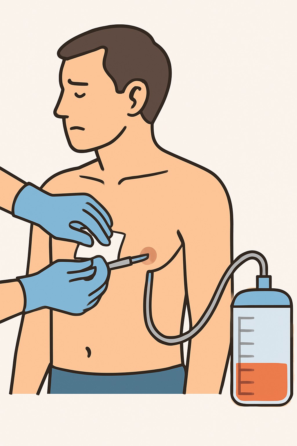
Chest tube insertion (thoracostomy) places a flexible tube through the chest wall into the pleural space, the area between the lung and the chest wall. It removes air, blood, pus, or fluid so the lung can expand and breathe more easily. Thoracostomy means making an opening in the chest to access this space. The procedure is usually done in a hospital or emergency setting with local anesthesia and sometimes sedation. The tube connects to a drainage system, which may use suction or a water seal, and imaging such as ultrasound or X-ray is often used to guide placement and check position.
Also known as: Tube thoracostomy, Chest drain, Pleural drain
Preparation & Next Steps
Everything you need to know before and after your procedure
Before Care
- Expect imaging (chest X-ray or ultrasound) to confirm the problem and guide the plan
- Share a current list of medicines, including blood thinners and over-the-counter products
- Tell the team about allergies, especially to local anesthetics (like lidocaine), antiseptics, or latex
- You may be asked not to eat or drink for several hours if sedation is planned
- Bring photo ID and insurance information and be ready to review and sign consent forms
- An IV line and basic blood tests may be done to check bleeding and infection risk
- Discuss pain control and sedation options available at the facility
- Wear loose clothing and leave valuables at home when possible
- Arrange a ride home if you are not being admitted after the procedure
- Follow any additional instructions from the care team or facility
After Care
- Keep the drainage system below chest level and ensure the tubing is not kinked or pulled
- Do not clamp the tube unless the care team instructs you to do so
- Use deep-breathing exercises and cough regularly to help the lung re-expand
- Walk and change position as allowed to reduce complications like pneumonia or blood clots
- Keep the dressing clean and dry; notify staff if it becomes loose, wet, or soaked
- If asked, help track drainage amount and appearance so the team can decide next steps
- Take pain medicine and other prescribed medicines as directed by your clinician
- Attend follow-up imaging or checks to confirm tube position and lung expansion
- Contact your care team if you have fever, increasing shortness of breath, spreading redness, drainage with pus, new swelling under the skin, sudden change in drainage, or if the tube or connections come apart
- After tube removal, keep the site covered and dry as instructed and avoid soaking the area until healed
Clinical Information
Important medical details about this procedure
Indications
- Collapsed lung (pneumothorax), including traumatic or spontaneous
- Blood in the chest (hemothorax)
- Large or recurrent pleural effusion
- Infected pleural fluid (empyema)
- Postoperative drainage after chest or heart surgery
- Chylothorax (lymphatic fluid in the pleural space)
Alternatives
- Observation with oxygen for some small pneumothoraces
- Needle aspiration for selected pneumothoraces
- Thoracentesis to remove pleural fluid
- Small-bore catheter with one-way (Heimlich) valve in selected cases
- Video-assisted thoracoscopic surgery (VATS) for persistent air leak or loculated fluid
Risks
- Pain or discomfort at the insertion site
- Bleeding or injury to intercostal blood vessels
- Infection at the skin or in the pleural space
- Lung injury or persistent air leak
- Injury to diaphragm, liver, or spleen depending on the site
- Tube misplacement, blockage, or accidental removal
- Air under the skin (subcutaneous emphysema)
- Re-expansion pulmonary edema
- Scarring or numbness near the incision
Contraindications
- Few absolute contraindications in emergencies
- Uncontrolled bleeding disorder (coagulopathy)
- Infection at the planned insertion site
- Dense pleural adhesions or prior pleurodesis at the site
- Inability to cooperate without airway protection when sedation is unsafe
Recovery Timeline
What to expect during your recovery
Recovery depends on the reason for the tube and overall health. Many people are active while the tube is in place and increase activity after it is removed. Soreness around the insertion site can last for several days.
Typical Range
Same day
Return to Work
Same day
Recovery Milestones
Begin short walks with assistance as allowed while keeping the drainage system below chest level
Use deep-breathing exercises and coughing to keep lungs open
After tube removal, gradually return to usual activities as soreness improves
Frequently Asked Questions
Common questions and expert answers about this procedure
What does a chest tube do?
What does a chest tube do?
It drains air, blood, pus, or fluid from the pleural space so the lung can expand and breathe more easily.
Will I be awake for the procedure?
Will I be awake for the procedure?
Most chest tubes are placed with local anesthesia and sometimes sedation. If placed during surgery, it is done under general anesthesia.
Is chest tube insertion painful?
Is chest tube insertion painful?
You will receive numbing medicine, and sometimes sedation, but pressure and soreness are common. Pain medicine is typically used during recovery.
How long does a chest tube stay in?
How long does a chest tube stay in?
It depends on why it was placed and how your lung and drainage change over time. Tubes are removed when the air leak stops, drainage decreases, and imaging looks improved.
Can I move around with a chest tube?
Can I move around with a chest tube?
Yes. Walking and sitting up are often encouraged. The drainage system should stay below chest level and the tubing should not be kinked or pulled.
How is the chest tube removed?
How is the chest tube removed?
Removal is usually done at the bedside. After cleaning and loosening the dressing, the tube is pulled quickly and the site is covered with a tight dressing.
What complications should I watch for?
What complications should I watch for?
Contact the care team if you notice fever, worsening shortness of breath, increasing redness or pus at the site, new swelling under the skin, sudden drainage changes, or if the tube or connections come apart.
Will there be a scar?
Will there be a scar?
A small scar at the insertion site is common. Some people notice numbness or firmness in that area for a while.
References
Medical literature and sources