Laser Lithotripsy (Ureteroscopic stone removal) Urologic Surgery
Laser lithotripsy during ureteroscopy is a minimally invasive procedure to treat stones in the ureter or kidney.
Overview
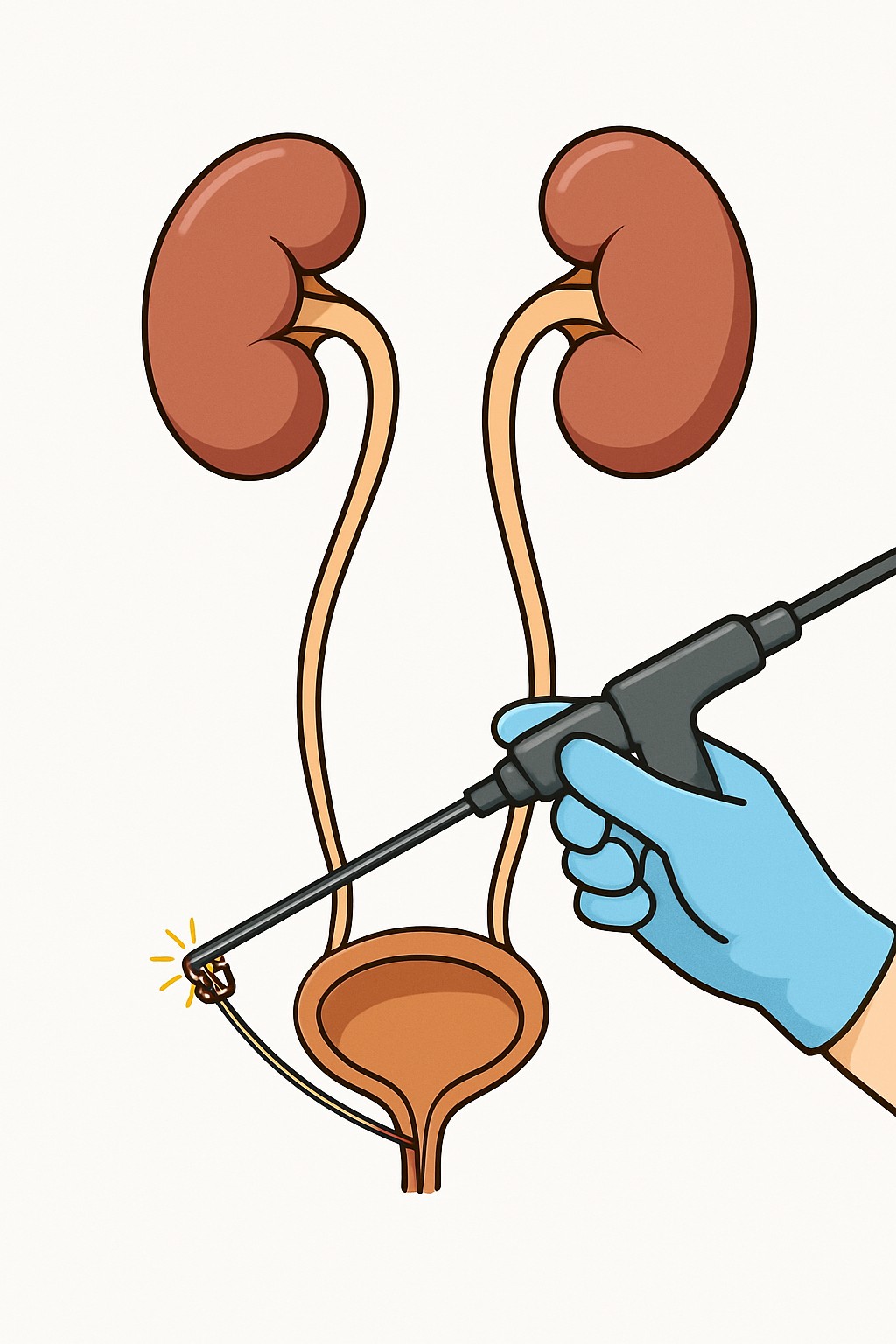
Laser lithotripsy during ureteroscopy is a minimally invasive procedure to treat stones in the ureter or kidney. A thin camera (ureteroscope) is passed through the urethra and bladder into the ureter or kidney to see the stone. A laser fiber breaks the stone into small pieces. Fragments may be removed with a tiny basket or left to pass naturally. A temporary soft tube called a ureteral stent is often placed to keep urine flowing and reduce swelling.
Also known as: Ureteroscopy with laser lithotripsy, Ureteroscopic stone removal, URS with laser, Ureteroscopy and laser fragmentation
Preparation & Next Steps
Everything you need to know before and after your procedure
Before Care
- Complete any requested urine test to check for infection before the procedure
- Follow instructions about holding blood thinners and supplements that increase bleeding risk
- Do not eat or drink for the directed period before anesthesia
- Arrange a responsible adult to drive you home after anesthesia
- Share allergies, implanted devices, and prior reactions to anesthesia with the care team
- Bring a current list of medicines and any recent imaging reports
- Plan for the possibility of a temporary ureteral stent after the procedure
- Wear comfortable, loose clothing and leave valuables at home
- Confirm check-in time, location, and any pre-op lab or paperwork needs
After Care
- Expect mild burning with urination and pink or red urine for 1–3 days
- Use a urine strainer if provided to collect stone fragments for analysis
- Follow the medicine plan provided by your clinician and pharmacy
- Stay active with light walking and avoid strenuous activity until you feel ready
- If a stent was placed, learn common stent symptoms (frequency, urgency, flank discomfort) and the removal plan
- Drink fluids as allowed to help flush small fragments
- Schedule and attend follow-up to review results, imaging, and stent removal timing if applicable
- Contact the clinic for fever of 100.4 F (38 C) or higher, inability to urinate, worsening flank pain, large blood clots, or foul-smelling urine
- Keep track of symptoms and any side effects to discuss at follow-up
Clinical Information
Important medical details about this procedure
Indications
- Painful ureteral or kidney stones
- Stone blocking urine flow (obstruction) or causing hydronephrosis
- Infection risk from an obstructing stone
- Failure of watchful waiting or shock wave treatment
- Stones not suitable for shock wave treatment
- Need for stone analysis to guide prevention
Alternatives
- Watchful waiting with fluids and pain control for small stones
- Medical expulsive therapy (alpha-blocker medicines that relax the ureter)
- Extracorporeal shock wave lithotripsy (ESWL)
- Percutaneous nephrolithotomy (PCNL) for larger or complex stones
- Temporary ureteral stent or nephrostomy tube to relieve blockage
- Open or laparoscopic surgery in rare cases
Risks
- Bleeding or blood in the urine
- Infection or sepsis
- Ureter injury, perforation, or later scar (stricture)
- Pain or discomfort from a ureteral stent
- Incomplete stone removal and need for repeat procedure
- Urinary retention or difficulty urinating
- Anesthesia-related risks
Contraindications
- Active, untreated urinary tract infection
- Uncorrected bleeding disorder or medicines that strongly increase bleeding risk
- Inability to tolerate anesthesia
- Severe urethral or ureteral narrowing that prevents safe scope passage
Recovery Timeline
What to expect during your recovery
Most people go home the same day and resume light activities within a few days. Discomfort usually improves over several days, especially if a stent is present. Full recovery varies with stone size, location, and whether a stent was placed.
Typical Range
2–7 days
Return to Work
2–7 days
Recovery Milestones
Walk short distances and urinate on your own after anesthesia wears off
Light activities at home; many people tolerate desk work
Pass small stone fragments; urine color gradually clears
Ureteral stent removal is often scheduled during this window, if one was placed
Increase activity as comfortable; avoid heavy lifting until cleared by the care team
Frequently Asked Questions
Common questions and expert answers about this procedure
What happens during laser lithotripsy with ureteroscopy?
What happens during laser lithotripsy with ureteroscopy?
A thin scope is passed through the urethra and bladder into the ureter or kidney. A laser breaks the stone, and pieces are either removed with a small basket or left to pass naturally. A temporary stent may be placed.
How long does the procedure take and is it outpatient?
How long does the procedure take and is it outpatient?
Many cases take 30–90 minutes, depending on stone size and location. It is usually an outpatient procedure with same-day discharge.
Will I be asleep for the procedure?
Will I be asleep for the procedure?
General anesthesia is commonly used so you are asleep and comfortable. The anesthesia plan is set by the anesthesia team.
Will I need a ureteral stent?
Will I need a ureteral stent?
A stent is often placed to keep the ureter open and help urine drain while swelling settles. It can cause urgency, frequency, or flank discomfort until removal.
What will recovery feel like?
What will recovery feel like?
It is common to have burning with urination, bladder spasms, and blood-tinged urine for a few days. Symptoms are often more noticeable if a stent is in place.
What are the risks?
What are the risks?
Risks include bleeding, infection, injury to the ureter, need for a stent, and the possibility of needing another procedure if fragments remain.
Can all stones be treated this way?
Can all stones be treated this way?
Most ureteral and many kidney stones can be treated. Very large or complex stones may be managed with percutaneous nephrolithotomy or staged procedures.
What if parts of the stone remain?
What if parts of the stone remain?
Small fragments may pass over days to weeks. Follow-up imaging checks for residual pieces. Sometimes a second procedure is planned.