IVC filter removal (vena cava filter retrieval) Vascular Surgery
IVC filter removal is a minimally invasive procedure to take out a small metal filter from the inferior vena cava, the large vein that carries blood from the lower body to the heart.
Overview
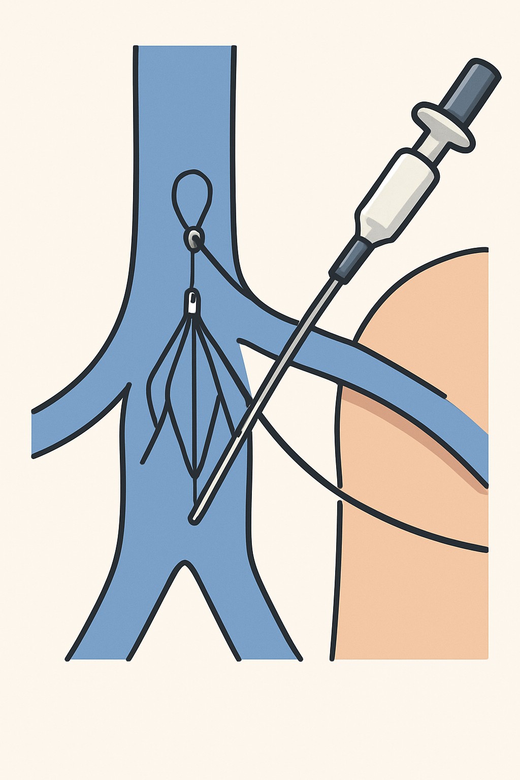
IVC filter removal is a minimally invasive procedure to take out a small metal filter from the inferior vena cava, the large vein that carries blood from the lower body to the heart. The filter is often placed to catch blood clots traveling from the legs and pelvis. When the risk of pulmonary embolism has passed or if the filter is causing problems, a specialist (often an interventional radiologist) may remove it. Using imaging guidance, the clinician threads a thin tube through a neck or groin vein, catches the filter with a snare, and withdraws it through the catheter.
Also known as: Inferior vena cava filter removal, IVC filter retrieval, Vena cava filter removal
Preparation & Next Steps
Everything you need to know before and after your procedure
Before Care
- Bring a list of all medicines and supplements, especially blood thinners, and share prior reactions to contrast dye or anesthesia
- Confirm which vein access is planned (neck or groin) and arrange comfortable clothing for easy access
- Follow any instructions about not eating or drinking if sedation is expected
- Arrange a responsible adult to drive you home if sedation or anesthesia is used
- Share recent imaging and procedure reports related to the filter placement and any follow-up scans
- Tell the care team about bleeding disorders, implanted devices, and allergies
- Ask how current conditions (such as pregnancy or kidney disease) may affect imaging contrast use
- Complete any requested labs or imaging (for example, kidney function tests or vein ultrasound) before the visit
- Plan time for check-in, consent, and recovery observation after the procedure
- Bring your insurance card, ID, and a payment method for any copay
After Care
- Keep the access site clean and dry as instructed; avoid soaking the area until cleared by your care team
- Expect mild soreness or bruising at the site; use simple comfort measures as advised by your clinician
- Limit heavy lifting and strenuous activity for a short period; resume normal light activity as you feel able
- Drink fluids if you received contrast dye, unless you were given different instructions
- Check the access site daily for redness, warmth, swelling, drainage, or increasing pain
- Contact your care team if you have chest pain, shortness of breath, fainting, fever, or bleeding that does not stop with gentle pressure
- Take prescribed medicines as directed by your clinician and keep your medication list updated
- Attend scheduled follow-up to review results and any imaging of the vena cava and leg veins
- Ask when you can return to work, driving, exercise, and sports based on how you feel and the type of work you do
- Save your discharge paperwork; it will list the filter type, removal details, and next steps
Clinical Information
Important medical details about this procedure
Indications
- Filter is no longer needed to prevent pulmonary embolism
- Filter-related problems such as pain, tilt, movement, fracture, or vein wall penetration
- Filter or vein clotting (occlusion) linked to the device
- Preparation for long-term anticoagulation without a filter
- Following safety guidance to remove a retrievable filter when appropriate
Alternatives
- Leave the filter in place with periodic imaging and follow-up
- Medical management with anticoagulation medicines when appropriate
- Advanced retrieval at a specialized center if standard removal is not possible
- Open surgical removal in rare situations when endovascular methods fail
Risks
- Bleeding or bruising at the vein access site
- Infection
- Allergic reaction to contrast dye
- Damage to the vein or nearby structures
- Blood clots or pulmonary embolism during or after the procedure
- Filter fracture or incomplete removal
- Need for a second procedure or open surgery in rare cases
Contraindications
- Active infection at the planned access site
- Uncontrolled bleeding or severe bleeding risk
- Severe allergy to iodinated contrast without a plan for alternatives
- Poor kidney function that limits safe use of contrast, if alternative imaging is not available
- Large clot trapped in the filter that cannot be safely managed
Recovery Timeline
What to expect during your recovery
Most people go home the same day. Soreness at the access site usually improves over a few days. Many return to normal routines within about a week, depending on activity level and sedation.
Typical Range
1–7 days
Return to Work
1–3 days
Recovery Milestones
Walk short distances and do light indoor activities
Increase routine activities as soreness improves
Avoid heavy lifting and high-intensity exercise until cleared
Resume usual activities if the access site is healed and you feel well
Frequently Asked Questions
Common questions and expert answers about this procedure
What is an IVC filter removal?
What is an IVC filter removal?
It is a procedure to take out a filter from the large vein in your abdomen using a catheter and imaging guidance.
Why are filters removed?
Why are filters removed?
They are removed when the risk of pulmonary embolism has passed or if the filter is causing problems such as movement, tilt, pain, or vein blockage.
How is the procedure performed?
How is the procedure performed?
A thin tube is inserted into a neck or groin vein. Using X-ray guidance, the clinician snags the filter with a snare and pulls it into the tube to remove it.
Will I be awake?
Will I be awake?
You may get local numbing medicine plus sedation to help you relax. The approach depends on the center and your health history.
How long does it take?
How long does it take?
Many removals take under an hour, but time varies based on filter type, position, and any scar tissue.
What if the filter cannot be removed?
What if the filter cannot be removed?
The team may try advanced techniques or refer to a specialized center. If removal is not safe, the filter may be left in place with follow-up.
What tests are needed before removal?
What tests are needed before removal?
Common steps include reviewing prior images, checking kidney function if contrast is used, and imaging to locate the filter and nearby clots.
Are there long-term effects after removal?
Are there long-term effects after removal?
Most people recover fully. Follow-up may include imaging to confirm the vein is open and to monitor for new clots if you are at risk.
References
Medical literature and sources